Incidental finding of impacted supernumerary teeth in cone-beam computed tomography 3D
Abstract
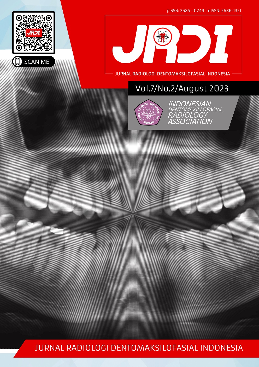
Objectives: To evaluate the value of cone-beam CT (CBCT) in the diagnosis and orientation of impacted supernumerary teeth in the dental arches.Case Report: A 50 year old man came to the Udayana University Hospital with the chief complaint of missing bilateral posterior teeth of the mandible and wanted to make denture. Before the treatment begin, a panoramic radiographic examination was performed. The panoramic image shows an impacted supernumerary teeth on the inferior from lower right premolar. Due to the inability to determine the precise position of those teeth within the mandible and the possible vital structures surrounding, a CBCT imaging was taken. Examination of the images from the CBCT shows an impacted supernumerary teeth in the area of teeth 44 and 46.
Conclusion: The position of the supernumerary teeth is varied in the mandible, and often causes permanent dentition complications. In this case CBCT imaging two supernumerary teeth were found in the tooth area 44-45 and 45-46. apical of the two germ teeth appear to be fully formed. Supernumerary crowns between teeth 44-45 inclined buccally & between teeth 45-46 lingually. CBCT is crucial for exact localization, for treatment planning, and for the surgical approach in cases of multiple supernumerary teeth.
References
Acharya S. Supernumerary teeth in maxillary anterior region : Report of three cases and their management. Int J Sci Study. 2015;3(3):122-7.
AL-Omar AF, Dakrory UAERE. Cone beam computed tomography for evaluation of impacted supernumerary teeth, Oral Health Care J. 2017;2(4):1-2.
Ahmad M, Jenny J, Downie M. Application of cone beam computed tomography in oral and maxillofacial surgery. Australian Dental Journal. 2012;57(Suppl 1):82–94.
Brauer HU. Case report: Non-syndromic multiple supernumerary teeth localized by cone beam computed tomography. Eur Arch Paediatr Dent. 2010;11(1):41-3.
Walker L, Enciso R, Mah J. Three-dimensional localization of maxillary canines with cone-beam computed tomography. Am J Orthod Dentofacial Orthop. 2015;128(4):418-23.
Sharma G, Nagra A, Singh G, Nagpal A, Soin A, Bhardwaj V. An Erupted Dilated Odontoma: A Rare Presentation. Case Rep Dent. 2016;2016:9750947.
Jung Y, Kim J, Cho B. The effects of impacted premaxillary supernumerary teeth on permanent incisors. Imaging Sci Dent. 2016;46:251-8.
White SC, Pharoah MJ. Oral Radiology and Interpretation. 7th ed. Canada: Elsevier Inc.; 2014.
Agarwal A, Shrivastava K, Tiwari P, PJaju P. CBCT Findings in Cleidocranial Dysplasia. Sch J Dent Sci. 2020;07(01): 19–23.
Toureno L, Park JH, Cederberg RA, Hwang EH, Shin JW. Identification of Supernumerary Teeth in 2D and 3D: Review of Literature and a Proposal. J Dent Educ. 2013;77(1):43–50
Dalessandri D, Laffranchi L, Tonni I, Zotti F, Piancino MG, Paganelli C, Bracco P. Advantages of cone beam computed tomography (CBCT) in the orthodontic treatment planning of cleidocranial dysplasia patients: a case report. Head Face Med. 2011;7:6.
Omami G. Multiple unerupted and supernumerary teeth in a patient with cleidocranial dysplasia. Radiol Case Reports. 2018;13(1):118–20.
Yusof WZ. Non-syndromal multiple supernumerary teeth: Literature review. J Can Dent Assoc. 1990;56(2):147-9.
Mallya SM, Lam EWN. White and Pharoah’s Oral Radiology Principles and Interpretation. 8th edition. Elsevier; 2019.
Rajab LD, Hamdan MA. Supernumerary teeth: Review of the literature and a survey of 152 cases. Int J Pediatr Dent. 2013;12:244-54.
Mitchell L. An introduction to orthodontics. 3rd ed. Oxford: Oxford University Press; 2007. p.24-7.
Published
2023-08-31
How to Cite
SUKMADEWI, Putri Marina; AGUNG, Anak Agung Gde Dananjaya.
Incidental finding of impacted supernumerary teeth in cone-beam computed tomography 3D.
Jurnal Radiologi Dentomaksilofasial Indonesia (JRDI), [S.l.], v. 7, n. 2, p. 79-82, aug. 2023.
ISSN 2686-1321.
Available at: <http://jurnal.pdgi.or.id/index.php/jrdi/article/view/1059>. Date accessed: 09 feb. 2026.
doi: https://doi.org/10.32793/jrdi.v7i2.1059.
Section
Case Report

This work is licensed under a Creative Commons Attribution-NonCommercial-NoDerivatives 4.0 International License.


