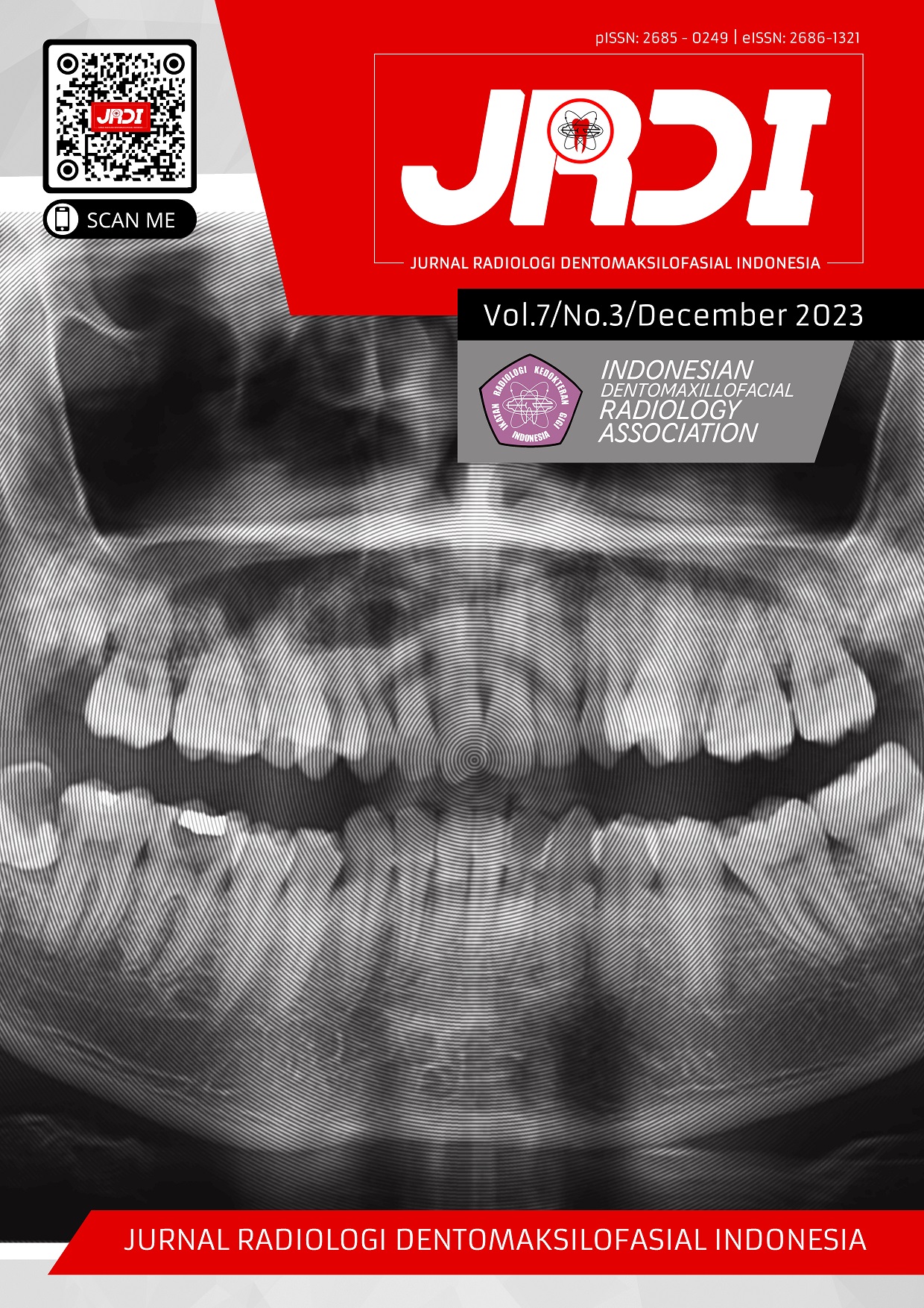
Age-related mandibular condyle morphological variations: a panoramic radiography study at RSGMP Universitas Airlangga
Abstract
Objectives: This research aims to find out the variations of the mandibular condyle shape based on age on panoramic radiography.Materials and Methods: This study uses secondary data from 200 digital panoramic radiographs of patients at the dental hospital (RSGM) of Universitas Airlangga aged 20-65 years in 2019, 2020, and 2021, which have met the inclusion and exclusion criteria. Data is presented in the form of tables and graphs with simple statistical calculations, and reliability tests were carried out with intraclass correlation (ICC) methods using SPSS.
Results: There are variations of the condyle shape in five age groups, the age I (20-25 Years), age II (26-35 years), age III (36-45 years), age IV (46-55 years), age V (56-65 years).
Conclusion: There are condyle shape variabilities in several age groups. In age I (20-25 years), age II (26-35 years), age III (36-45 years), and age IV (46-55 years) the most variation of the condyle shape is rounded, at age V (56-65 years) the most variation of the condyle shape is rounded and pointed. Furthermore, the morphology of the condylar structures may exhibit variances and are not consistently uniform.
References
Mathew AL, Sholapurkar AA, Pai KM. Condylar Changes and Its Association with Age, TMD, and Dentition Status: A Cross-Sectional Study. Int J Dent. 2011;2011:413639.
Alomar X, Medrano J, Cabratosa J, Clavero JA, Lorente M, Serra I, Monill JM, Salvador A. Anatomy of the temporomandibular joint. Semin Ultrasound CT MR. 2007;28(3):170-83.
Hegde S, Praveen BN, Shetty SR. Morphological and Radiological Variations of Mandibular Condyles in Health and Diseases: A Systematic Review. Dentistry. 2013;3(1):1–5.
Singh B, Kumar NR, Balan A, Nishan M, Haris PS, Jinisha M, Denny CD. Evaluation of Normal Morphology of Mandibular Condyle: A Radiographic Survey. J Clin Imaging Sci. 2020 17;10:51.
Vahanwala S, Pagare S, Gavand K, Roy C. Evaluation of condyle morphology using panoramic radiography. Journal of Advanced Clinical & Research Insights. 2016;3:5–8.
Song J, Cheng M, Qian Y, Chu F. Cone-beam CT evaluation of temporomandibular joint in permanent dentition according to Angle's classification. Oral Radiol. 2020 Jul;36(3):261-6.
Ishibashi H, Takenoshita Y, Ishibashi K, Oka M. Age-related changes in the human mandibular condyle: a morphologic, radiologic, and histologic study. J Oral Maxillofac Surg. 1995 Sep;53(9):1016-23; discussion 1023-4.
Nurfaidah WR. Gambaran morfologi kepala kondilus mandibula ditinjau dari radiograf panoramik berdasarkan usia [Skripsi]. Bandung: Universitas Padjajaran Repository; 2019.
Valladares Neto J, Estrela C, Bueno MR, Guedes OA, Porto OCL, Pécora JD. Mandibular condyle dimensional changes in subjects from 3 to 20 years of age using Cone-Beam Computed Tomography: A preliminary study. Dental Press Journal of Orthodontics. 2010;15(5):172–81.
White SC, Pharoah MJ. Oral Radiology Principles and Interpretation 7th Edition. Elsevier Inc; 2014.
Ismunarti DH, Zainuri M, Sugianto DN, Saputra SW. Pengujian Reliabilitas Instrumen Terhadap Variabel Kontinu Untuk Pengukuran Konsentrasi Klorofil- A Perairan. Buletin Oseanografi Marina. 2020;9(1):1–8.
Oliveira-Santos C, Bernardo, RT, Capelozza ALA. Mandibular condyle morphology on panoramic radiographs of asymptomatic temporomandibular joints. International Journal of Dentistry. 2009;8(3):114–8.
Whyte A, Boeddinghaus R, Bartley A, Vijeyaendra R. Imaging of the temporomandibular joint. Clinical Radiology. 2021:76(1);21-35.
Tecco S, Saccucci M, Nucera R, Polimeni A, Pagnoni M, Cordasco G, Festa F, Iannetti G. Condylar volume and surface in Caucasian young adult subjects. BMC Med Imaging. 2010;10:28.
Kurnia SI, Himawan LS, Odang RW. Correlation between Chewing Preference and Condyle Asymmetry in Patients with Temporomandibular Disorders. Journal of Physics: Conference Series. 2018;1073(3):032014.
Published
2023-12-31
How to Cite
MULYANI, Sri Wigati Mardi et al.
Age-related mandibular condyle morphological variations: a panoramic radiography study at RSGMP Universitas Airlangga.
Jurnal Radiologi Dentomaksilofasial Indonesia (JRDI), [S.l.], v. 7, n. 3, p. 89-94, dec. 2023.
ISSN 2686-1321.
Available at: <http://jurnal.pdgi.or.id/index.php/jrdi/article/view/1081>. Date accessed: 25 feb. 2026.
doi: https://doi.org/10.32793/jrdi.v7i3.1081.
Section
Original Research Article

This work is licensed under a Creative Commons Attribution-NonCommercial-NoDerivatives 4.0 International License.


