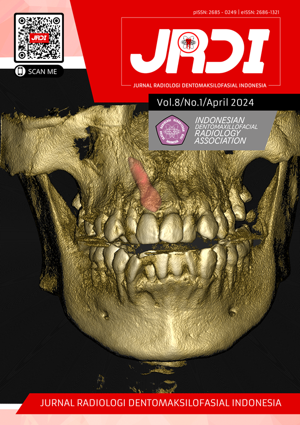
Enhancing the diagnosis of sublingual sialolith using CBCT: a case report
Abstract
Objectives: This case report was created to provide further insight into the use of CBCT in detecting sialolith lesions.Case Report: A 27-year-old female patient was referred to the radiology department of Padjadjaran University Dental Hospital for CBCT radiograph examination related to complaints of swelling in the right lingual area of the mandible and pain. The results of the radiograph examination analysis showed irregular, well-defined radiopaque, in the lingual area of regio 44-45, measuring 34.31 mm2, and the lesion was not associated with the mandible. Intra-oral examination revealed irregular swelling and the same color as the lingual mucosa of regio 44-45. The analysis showed that the lesion was located in the salivary duct of the sublingual area and the patient was diagnosed with sublingual sialolithiasis dextra.
Conclusion: CBCT analysis can be used to accurately identify the position, quantity, and morphology of sialoliths and interpret their three-dimensional positioning in relation to adjacent structures.
References
Tassoker M, Ozcan S. Two Cases of Submandibular Sialolithiasis Detected by Cone Beam Computed Tomography. IOSR Journal of Dental and Medical Sciences. 2016;15(08):124-9.
Thomas WW, Douglas JE, Rassekh CH. Accuracy of Ultrasonography and Computed Tomography in the Evaluation of Patients Undergoing Sialendoscopy for Sialolithiasis. Otolaryngology - Head and Neck Surgery (United States). 2017;156(5):834-9.
Kuruvila VE, Bilahari N, Kumari B, James B. Submandibular sialolithiasis: Report of six cases. J Pharm Bioallied Sci. 2013;5(3):240-2.
Kim JH, Aoki EM, Cortes ARG, Abdala-Júnior R, Asaumi J, Arita ES. Comparison of the diagnostic performance of panoramic and occlusal radiographs in detecting submandibular sialoliths. Imaging Sci Dent. 2016;46(2):87.
Abdel-Wahed N, Amer ME, Abo-Taleb NSM. Assessment of the role of cone beam computed sialography in diagnosing salivary gland lesions. Imaging Sci Dent. 2013;43(1):17-23.
Pachisia S, Mandal G, Sahu S, Ghosh S. Submandibular Sialolithiasis: A Series of Three Case Reports with Review of Literature. Clin Pract. 2019;9(1):1119.
Lim EH, Nadarajah S, Mohamad I. Giant Submandibular Calculus Eroding Oral Cavity Mucosa. Oman Med J. 2017;32(5):432-5.
Gadve V, Mohite A, Bang K, Shenoi S. Unusual giant sialolith of Wharton’s duct. Indian J Dent. 2016;7(3):162.
Jadu F, Yaffe M, Lam E. A comparative study of the effective radiation doses from cone beam computed tomography and plain radiography for sialography. Dentomaxillofacial Radiology. 2010;39(5):257-63.
Demidov V, Khrulenko S. Sialoliths of Submandibular Gland and Wharton’s Duct: Orthopantomography. Journal of Diagnostics and Treatment of Oral and Maxillofacial Pathology. 2021;5(7):77-86.
Song Y Bin, Jeong HG, Kim C, et al. Comparison of detection performance of soft tissue calcifications using artificial intelligence in panoramic radiography. Sci Rep. 2022;12(1):19115.
Veniaminivna Kolomiiets S, Oleksandrivna Udaltsova K, Andriivna Khmil T, Mykolaiivna Yelinska A, Anatoliivna Pisarenko O, Ihorivna Shynkevych V. Difficulties in Diagnosis of Sialolithiasis: A Case Series. Bull Tokyo Dent Coll. 2018;59(1):53-8.
van der Meij EH, Karagozoglu KH, de Visscher JGAM. The value of cone beam computed tomography in the detection of salivary stones prior to sialendoscopy. Int J Oral Maxillofac Surg. 2018;47(2):223-7.
Yajima A, Otonari-Yamamoto M, Sano T, et al. Cone-beam CT (CB Throne) Applied to Dentomaxillofacial Region. Bull Tokyo Dent Coll. 2006;47(3):133-41.
Cassetta M, Stefanelli L, Carlo S Di, Pompa G. The Accuracy of CBCT in Measuring Jaws Bone Densit. Pierre Robine Sequence Orthodontic Approach View Project Retrograde Peri-Implatitis View Project.; 2012. https://www.researchgate.net/publication/232721119
Santos JO, Da Silva Firmino B, Carvalho MS, et al. 3D reconstruction and prediction of sialolith surgery. Case Rep Dent. 2018;2018.
AlMadi DM, Al-Hadlaq MA, AlOtaibi O, Alshagroud RS, Al-Ekrish AA. Accuracy of mean grey density values obtained with small field of view cone beam computed tomography in differentiation between periapical cystic and solid lesions. Int Endod J. 2020;53(10):1318-26.
Costan VV, Ciocan-Pendefunda CC, Sulea D, Popescu E, Boisteanu O. Use of Cone-Beam Computed Tomography in Performing Submandibular Sialolithotomy. Journal of Oral and Maxillofacial Surgery. 2019;77(8):1656.e1-1656.e8.
Dreiseidler T, Ritter L, Rothamel D, Neugebauer J, Scheer M, Mischkowski RA. Salivary calculus diagnosis with 3-dimensional cone-beam computed tomography. Oral Surgery, Oral Medicine, Oral Pathology, Oral Radiology, and Endodontology. 2010;110(1):94-100.
Published
2024-04-30
How to Cite
PUTRA, Dimas Satria; EPSILAWATI, Lusi.
Enhancing the diagnosis of sublingual sialolith using CBCT: a case report.
Jurnal Radiologi Dentomaksilofasial Indonesia (JRDI), [S.l.], v. 8, n. 1, p. 29-32, apr. 2024.
ISSN 2686-1321.
Available at: <http://jurnal.pdgi.or.id/index.php/jrdi/article/view/1114>. Date accessed: 25 feb. 2026.
doi: https://doi.org/10.32793/jrdi.v8i1.1114.
Section
Case Report

This work is licensed under a Creative Commons Attribution-NonCommercial-NoDerivatives 4.0 International License.


