CBCT-3D radiographic analysis of an infected radicular cyst of the upper jaw: a case report
Abstract
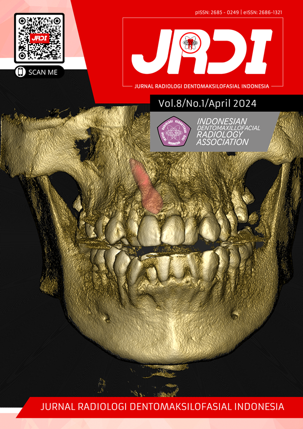
Objectives: The purpose of this case report is to find out the function of CBCT that can analyze in detail a radicular cyst case where CBCT has the ability to determine the density of a lesion. An overview of the density pattern of a radicular cyst can help determine the lesion and its margins.Case Report: : A 40-year-old patient complained of swelling in the palate that had been felt for about a year with a history of trauma due to an accident fifteen years ago. The patient underwent CBCT examination with the results of a hypodense area compared to the surrounding bone in the maxillary anterior area involving teeth 11,21,22 to 23 accompanied by a discontinuity of the cortical plate of the palatal bone and extending to involve the base of the nasal cavity. Part of the lesion is clearly demarcated, while the other side is diffuse. The suspected radiodiagnosis of the case was a moderately aggressive radicular cyst with a differential diagnosis of an odontogenic tumor.
Conclusion: The final diagnosis of this case was a radicular cyst with chronic granulomatous inflammation, based on a combination of radiographic findings, clinical findings, and histopathological examination.
References
Althaf S, Hussaini N, Srirekha A, Santhosh L. The role of cone-beam computed tomography in evaluation of an extensive radicular cyst of the maxilla. Journal of Restorative Dentistry and Endodontics. 2021;1(1):30-3.
Latoo S, Shah AA, Jan SM, Qadir S, Ahmed I, Purra AR et al. Radicular Cyst. JK Science. 2009;11(4):187-9.
Singh J, Bhagat S, Aneja A, Sharma R, Kaur M. Management of Radicular Cyst- A Case Report. Annal Cas Rep Clin Stud (ACRCS). 2023;2(3):1-5.
Viggness A, Priyaa S, Thangavelu RP, Fenn SM, Mohan KR. The Three-Dimensional Evaluation of Radicular Cyst by Cone-Beam Computed Tomography. Cureus. 2023;15(3): e36162.
Brave D, Madhusudan AS, Ramesh G, Brave VR. Radicular cyst of anterior Maxilla. USA: International Journal of Dental Clinics. 2011:3(4):16-17.
Koju S, Chaurasia NK, Marla V, Deepa N, Poudel P. Radicular Cyst of The anterior Maxilla: An Insight into the Most Common Inflammatory Cyst of the Jaws. Journal of Dental Research and Review. 2019;6(1):26-9.
Velasco I, Vahdani S, Nuñez N, Ramos H. Large Recurrent Radicular Cyst in Maxillary Sinus: A Case Report. Int J Odontostomat. 2017;11(1):101-5.
Andresen AKH, Jonsson MV, Sulo G, Thelen DS, Shi XQ. Radiographic features in 2D imaging as predictors for justified CBCT examinations of canine-induced root resorption. Dentomaxillofac Radiol. 2022;51(1):20210165.
Kumar A, Singh N, Singh S, Talukdar M, Kisave PN, Saba I. Occurrence of radicular cyst in anterior maxilla of adolescent patient: A case report and its management. International Journal of Applied Dental Sciences. 2019; 5(1):20-2.
Pei J, Zhao S, Chen H, Wang J. Management of radicular cyst associated with primary teeth using decompression: a retrospective study. BMC Oral Health. 2022;22(1):560.
Prativi SA, Pramatika B. Gambaran Karakteristik Kista Radikular Menggunakan Cone Beam Computed Tomography (CBCT): Laporan Kasus. B-Dent: Jurnal Kedokteran Gigi Universitas Baiturrahmah. 2019;6(2):105-10.
Kumaravelu R, Jude NJ, Sathyanarayanan R. Radicular Cyst: A Case Report. J Sci Dent. 2021;11(1):23–5.
Pratyusha MV, Nadig P, Jayalakshmi KB, Math S. CBCT assessment of healing of a large radicular cyst treated with enucleation followed by PRF and osseograft placement: A case report. Saudi J Oral Dent Res. 2017;2(3):72-5.
Rajendran R, Sivapathasundharam B. Shafer’s Textbook of Oral Pathology. 6th ed. New Delhi: Elsevier; 2009. p.487-90.
Pratyusha MV, Nadig P, Jayalakshmi KB, Math S. CBCT assessment of healing of a large radicular cyst treated with enucleation followed by PRF and osseograft placement: A case report. Saudi J Oral Dent Res 2017;2:72-5.
Deshmukh J, Shrivastava R, Bharath KP. Giant radicular cyst of the maxilla. BMJ Case Rep 2014;2014:bcr2014203678.
Kamburoglu K. Use of dentomaxillofacial cone beam computed tomography in dentistry. World J Radiol 2015;6:128-30.
Published
2024-04-30
How to Cite
PRAWIRA, Ade et al.
CBCT-3D radiographic analysis of an infected radicular cyst of the upper jaw: a case report.
Jurnal Radiologi Dentomaksilofasial Indonesia (JRDI), [S.l.], v. 8, n. 1, p. 33-36, apr. 2024.
ISSN 2686-1321.
Available at: <http://jurnal.pdgi.or.id/index.php/jrdi/article/view/1130>. Date accessed: 16 feb. 2026.
doi: https://doi.org/10.32793/jrdi.v8i1.1130.
Section
Case Report

This work is licensed under a Creative Commons Attribution-NonCommercial-NoDerivatives 4.0 International License.


