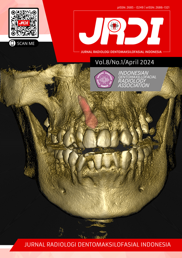
Case report: Detection of maxillary sinusitis with inverted impacted teeth using Cone-beam Computed Tomography
Abstract
Objectives: The aim of this case report is to describe radiographically the specific features of maxillary sinusitis on CBCT radiograph.Case Report: A 20-year-old female patient came to RSGM UNPAD with a consul letter from Oral Surgery specialist for a CBCT radiography examination to see impacted teeth. The results showed radiointermediate images in the maxillary sinus which showed thickening of the sinus mucosa and an inverted impacted teeth on the right maxillary.
Conclusion: Maxillary sinusitis could be assessed using extra oral radiography and CBCT. CBCT examination was used in determining the source of the lesion, the extent of the lesion, and the thickness of the maxillary sinus mucosa.
References
Krenz-Niedbała M, Łukasik S. Prevalence of chronic maxillary sinusitis in children from rural and urban skeletal populations in Poland. Int J Paleopathol. 2016;15:103–12.
TE K, B A, M B, İ Ö, HA B. Two Cases of Inverted Ectopic Teeth in Maxillary Sinus. 2016;1(3):3–5.
Serindere G, Bilgili E, Yesil C, Ozveren N. Evaluation of maxillary sinusitis from panoramic radiographs and cone-beam computed tomographic images using a convolutional neural network. Imaging Sci Dent. 2022;52:187–95.
Sinha A, Mishra A, Anusha, PM Sinha. Ectopic third molar in Maxillary sinus : A rare case report. Journal Indian Acad Oral Med Radiol. 2017;29(4):341-4.
K S, M I, M S, Y H, T K. Case Reports A Case of Impacted Tooth in the Maxillary Sinus : CT Findings. 2015;13(3):128–30.
Whaites E, Drage N. Essentials of Dental Radiography and Radiology. 6th ed. Elsevier; 2021.
SM M, EWN L. White and pharoah oral radiology : Principles and interpretation. Elsevier; 2014.
Bilginaylar K, Inancli HM, Kneebone M. Endoscopic and Intraoral Approach for Removal of an Ectopic Third Molar associated with a Dentigerous Cyst in the Maxillary Sinus: A Case Report. Journal of Clinical and Diagnostic Research. 2023;17(5):25–8.
Sugiharto AF, Studi P, Radiologi T, Terapan PS, Teknik J, Dan R, et al. PERANAN CONE-BEAM COMPUTED TOMOGRAPHY (CBCT) DALAM PENEGAKAN DIAGNOSIS AMELOBLASTOMA ( Literature Review ). 2022: 1-18.
Dhingra S, Gulati A. Teeth in Rare Locations with Rare Complications: An Overview. Indian Journal of Otolaryngology and Head and Neck Surgery. 2015;67(4):438–43.
Kara Mİ, Yanık S, Altan A, Öznalçın O, Ay S. Large Dentigerous Cyst in the Maxillary Sinus Leading To Diplopia and Nasal Obstruction: Case Report. J Istanb Univ Fac Dent. 2015;49(2):46.
Arora P, Nair MK, Liang H, Patel PB, Wright JM, Tahmasbi-Arashlow M. Ectopic teeth with disparate migration: A literature review and new case series. Imaging Sci Dent. 2023;53(3):229–38.
David CM, Kastala RK, Jayapal N, Majid SA. Imaging modalities for midfacial fractures. Trauma (United Kingdom). 2017;19(3):175–85.
Koenig LJ, Tamimi D, Perschbacher SE. Diagnostic Imaging: Oral and Maxillofacial. 2th ed. Elsevier; 2017.
Lin J, Wang C, Wang X, Chen F, Zhang W, Sun H, et al. Expert consensus on odontogenic maxillary sinusitis multi-disciplinary treatment. Int J Oral Sci. 2024;16(11):1-14.
Surenthar M, Srinivasan SV, Jimsha VK, Vineeth R. Clinical Dilemma and the Role of Cone-Beam Computed Tomography in the Diagnosis of an Unusual Presentation of Central Odontogenic Tumor-A Case Report. Indian Journal of Radiology and Imaging. 2021;31(3):782–8.
Suomalainen A, Pakbaznejad Esmaeili E, Robinson S. Dentomaxillofacial imaging with panoramic views and cone beam CT. Insights Imaging. 2015;6(1):1–16.
Alabdulwahid A. Cone beam computed tomography: still a blessing for maxillofacial imaging. J Dent Health Oral Disord Ther. 2021;12(2):33–9.
Nasseh I. Unusual Behavior of the Mandibular Canal Associated to a Dentigerous Cyst. J Dent Health Oral Disord Ther. 2015;2(2):53–5.
Elizabeth Mathen D, Susan Philip S, Rawther D, Padiyath D. Central Odontogenic Fibroma-A Deep Seated Lesion Unveiled. IOSR Journal of Dental and Medical Sciences (IOSR-JDMS) e-ISSN [Internet]. 2018;17(8):51–5. Available from: www.iosrjournals.org
Published
2024-04-30
How to Cite
DAMAYANTI, Merry Annisa et al.
Case report: Detection of maxillary sinusitis with inverted impacted teeth using Cone-beam Computed Tomography.
Jurnal Radiologi Dentomaksilofasial Indonesia (JRDI), [S.l.], v. 8, n. 1, p. 25-28, apr. 2024.
ISSN 2686-1321.
Available at: <http://jurnal.pdgi.or.id/index.php/jrdi/article/view/1175>. Date accessed: 25 feb. 2026.
doi: https://doi.org/10.32793/jrdi.v8i1.1175.
Section
Case Report

This work is licensed under a Creative Commons Attribution-NonCommercial-NoDerivatives 4.0 International License.


