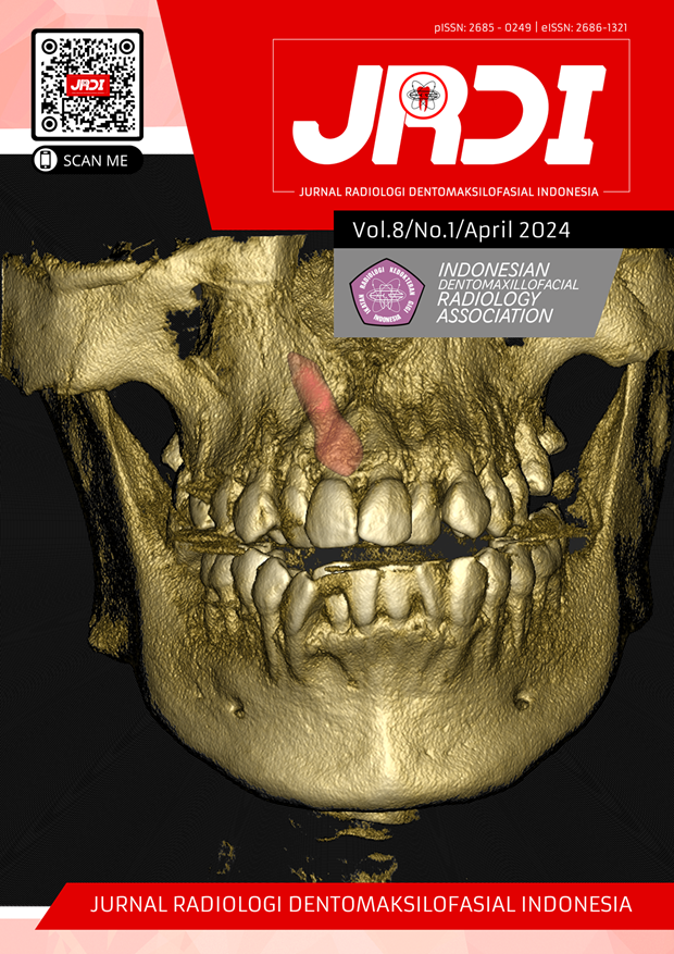
A novel approach of tongue cancer diagnostic imaging: a literature review
Abstract
Objectives: This review article is aimed to provide a scientific information about novel approach of tongue cancer diagnostic imaging based on evidence-based reputed published studies.Review: The databases used in this literature review are Google Scholar, PubMed and Elsevier. The research results were selected by title and abstract according to their relevance to the review topic, then the results were selected again based on the inclusion and exclusion criteria. A total of 13 literatures were reviewed. This review shows the diagnostic imaging is a useful tool for staging and management planning in tongue cancers. In this era of technological development, a novel diagnostic imaging technologies that can be used for the diagnosis of tongue cancers such us Ultra High-Frequency Ultrasound (UHFUS), Diffusion-Weighted Imaging MRI (DWI-MRI), Dynamic Contrast-Enhanced MRI (DCE-MRI), Optical Coherence Tomography (OCT), Positron Emission Tomography (PET), Endoscopic images, and the other noninvasive imaging methods like vital staining, autofluorescence, Narrow-Band Imaging (NBI), and in vivo confocal microscopy. Besides that, imaging of tongue cancer requires a multimodality imaging approach to obtain accurate information about pathological condition such as PET/MRI, FDG PET/CT, and SPECT-CT.
Conclusion: Each diagnostic imaging has limitations in imaging the patient's condition, so it can be used alone or in combination with one another to obtain accurate information about pathological conditions. FGD PET-CT and UHFUS reportedly provide a high level of sensitivity and specificity to diagnose and staging of tongue cancer.
References
Tagliabue M, Belloni P, Berardinis R De, Gandini S, Chu F, Zorzi S, et al. A systematic review and meta- analysis of the prognostic role of age in oral tongue cancer. J Wiley Cancer Med. 2021;10(2):2566–78.
Arrangoiz R, Cordera F, Caba D, Moreno E, León EL De, Muñoz M. Oral Tongue Cancer : Literature Review and Current Management. J Cancer Rep Rev. 2018;2(3):1–9.
Sarrión MG, Bagán J V, Jiménez Y, Margaix M, Marzal C. Utility Of Imaging Techniques In The Diagnosis Of Oral Cancer. J Cranio-Maxillo-Facial Surg. 2015;43(9):1–31.
Abhinaya LM, Muthukrishnan A. Clinical practice guidelines for radiographic assessment in management of oral cancer. J Adv Pharm Technol Res. 2022;13(4):248–51.
Widyaningrum R, Faisal A, Mitrayana M, Mudjosemedi M, Agustina D. Oral cancer imaging: the principles of interpretation on dental radiograph, CT, CBCT, MRI, and USG. Maj Kedokt Gigi Indones. 2018;4(1):1–10.
Nurrahman T, Adiantoro S, Rizki KA, Asnely F. Multidisciplinary Approach in the Treatment of Squamous Cell Carcinoma At Regio Glossus. Dentino J Kedokt Gigi. 2020;5(2):205–9.
Campbell BR, Netterville JL, Sinard RJ, Rohde SL, Langerman A, Kim YJ, et al. Early onset oral tongue cancer in the United States: a literature review. J Oral Oncol. 2020;87(12):1–7.
Singh D, Sahoo S, Srivastava D, Gupta V. Latest advancements in imaging of oral and maxillofacial neoplasm: A comprehensive review. J Oral Maxillofac Radiol. 2013;1(2):37–42.
Yang Z, Pan H, Shang J, Zhang J, Liang Y. Deep-Learning-Based Automated Identification and Visualization of Oral Cancer in Optical Coherence Tomography Images. Biomedicines. 2023;11(3):1–12.
Tang W, Wang Y, Yuan Y, Tao X. Assessment of tumor depth in oral tongue squamous cell carcinoma with multiparametric MRI: correlation with pathology. Eur Radiol. 2022;32(1):254–61.
Heo J, Lim JH, Lee HR, Jang JY, Shin YS, Kim D, et al. Deep learning model for tongue cancer diagnosis using endoscopic images. Sci Rep. 2022;12(1):1–10.
Aydos U, Cebeci S. Prognostic role of primary tumor metabolic-volumetric parameters of 18F-fluorodeoxyglucose positron emission tomography in tongue squamous cell carcinoma. J Heal Sci Med. 2023;6(1):181–7.
Novikov SN, Krzhivitskii PI, Radgabova ZA, Kotov MA, Girshovich MM, Artemyeva AS, et al. Single photon emission computed tomography-computed tomography visualization of sentinel lymph nodes for lymph flow guided nodal irradiation in oral tongue cancer. Radiat Oncol J. 2021;39(3):193–201.
Linz C, Brands RC, Herterich T, Hartmann S, Müller-Richter U, Kübler AC, et al. Accuracy of 18-F Fluorodeoxyglucose Positron Emission Tomographic/Computed Tomographic Imaging in Primary Staging of Squamous Cell Carcinoma of the Oral Cavity. JAMA Netw Open. 2021;4(4):1–12.
Izzetti R, Nisi M, Gennai S, Oranges T, Crocetti L, Caramella D, et al. Evaluation of depth of invasion in oral squamous cell carcinoma with ultra-high frequency ultrasound: A preliminary study. Appl Sci. 2021;11(16):1–9.
Iandelli A, Sampieri C, Marchi F, Pennacchi A, Carobbio ALC, Lovino Camerino P, et al. The Role of Peritumoral Depapillation and Its Impact on Narrow-Band Imaging in Oral Tongue Squamous Cell Carcinoma. J Cancers. 2023;15(4):1–14.
Ikeda Y, Suzuki T, Saitou H, Ogane S, Hashimoto K, Takano N, et al. Usefulness of fluorescence visualization-guided surgery for early-stage tongue squamous cell carcinoma compared to iodine vital staining. Int J Clin Oncol. 2020;25(9):1604–11.
Kanno M, Tsujikawa T, Narita N, Ito Y, Makino A, Imamura Y. Comparison of diagnostic accuracy between [ 18 F ] FDG PET / MRI and contrast ‑ enhanced MRI in T staging for oral tongue cancer. Ann Nucl Med. 2020;1(1):1–8.
Guo N, Zeng W, Deng H, Hu H, Cheng Z, Yang Z, et al. Quantitative dynamic contrast-enhanced MR imaging can be used to predict the pathologic stages of oral tongue squamous cell carcinoma. BMC Med Imaging. 2020;20(1):1–9.
Jeng MJ, Sharma M, Chao TY, Li YC, Huang SF, Chang LB, et al. Multiclass classification of autofluorescence images of oral cavity lesions based on quantitative analysis. J PLoS ONE. 2020;15(2):1–18.
Contaldo M, Lauritano D, Carinci F, Romano A, Stasio D Di, Lajolo C, et al. Intraoral confocal microscopy of suspicious oral lesions : a prospective case series. Int J Dermatol. 2020;1(1):1–9.
Dhoot NM, Hazarika S, Choudhury B, Kataki AC, Goswami H, Baruah R, et al. Pre-operative staging of carcinoma of tongue using Ultrasonography and Magnetic Resonance imaging. Am J Diagnostic Imaging. 2017;2(1):1–13.
Jerjes W, Hamdoon Z, Yousif AA, Al-Rawi NH, Hopper C. Epithelial tissue thickness improves optical coherence tomography’s ability in detecting oral cancer. Photodiagnosis Photodyn Ther. 2019;28(8):69–74.
Panzarella V, Buttacavoli F, Gambino A, Capocasale G, Fede O Di, Mauceri R, et al. Site-Coded Oral Squamous Cell Carcinoma Evaluation by Optical Coherence Tomography (OCT): A Descriptive Pilot Study. J Cancers. 2022;14(1):1–15.
Lohe V, Bhowate R, Parihar P, Kadu R, Sune R V, Mohod SC, et al. Evaluation of Lymph Nodes in Oral Squamous Cell Carcinoma by Diffusion ‑ Weighted Magnetic Resonance Imaging , Apparent Diffusion Coefficient Mapping , and Fast ‑ Spin Echo Magnetic Resonance Imaging. J Datta Meghe Inst Med Sci Univ |. 2022;1(1):1–5.
Chen CF, Peng SL, Lee CC, Lui CC, Huang HY, Chien CY. Dynamic contrast-enhanced magnetic resonance imaging in correlation with tongue cancer stages. Acta radiol. 2020;62(12):1–7.
Afonso PD, Mascarenhas V V. Imaging techniques for the diagnosis of soft tissue tumors. Reports Med Imaging. 2015;8(1):63–70.
Pałasz P, Adamski Ł, Górska-Chrząstek M, Starzyńska A, Studniarek M. Contemporary diagnostic imaging of oral squamous cell carcinoma – A review of literature. Polish J Radiol. 2017;82(1):193–202.
Romano A, Di Stasio D, Petruzzi M, Fiori F, Lajolo C, Santarelli A, et al. Noninvasive imaging methods to improve the diagnosis of oral carcinoma and its precursors: State of the art and proposal of a three-step diagnostic process. J Cancers. 2021;13(12):1–22.
Sreeshyla H, Sudheendra U, Shashidara R. Vital tissue staining in the diagnosis of oral precancer and cancer: Stains, technique, utility, and reliability. Clin Cancer Investig J. 2014;3(2):141–5.
Kawada K, Kawano T, Okada T, Yamaguchi K, Kawamura IY, Matsui T, et al. The usefulness of an intra-oropharyngeal u-turn method using trans-nasal endoscopy for detecting superficial squamous cell carcinoma of the base of the tongue. J Otolaryngol - ENT Res. 2017;8(2):422–7.
Vescovi P, Giovannacci I, Meleti M. Laser Applications and Autofluorescence. Eur J Mol Clin Med. 2020;7(3):140–50.
Huang T ta, Huang J shyun, Wang Y yun, Chen K chung, Wong T yiu, Yuan S shiou F, et al. Novel quantitative analysis of autofluorescence images for oral cancer screening. Oral Oncol. 2017;68(1):20–6.
Russo A, Reginelli A, Lacasella GV, Grassi E, Karaboue MAA, Quarto T, et al. Clinical Application of Ultra-High-Frequency Ultrasound. J Pers Med. 2022;12(10):1–11.
Izzetti R, Vitali S, Aringhieri G, Nisi M, Oranges T, Dini V, et al. Ultra-High Frequency Ultrasound, A Promising Diagnostic Technique: Review of the Literature and Single-Center Experience. Can Assoc Radiol J. 2021;20(10):1–15.
Saraniti C, Greco G, Verro B, Lazim NM, Chianetta E. Impact of narrow band imaging in pre-operative assessment of suspicious oral cavity lesions: A systematic review. Iran J Otorhinolaryngol. 2021;33(3):127–35.
Nikose P, Johar NS, Dosi T, Mahey P. In vivo microscopy : A non-invasive diagnostic tool. Int J Periodontol Implantol. 2022;6(4):226–9.
Vasudevan V, Mathew NS, Devaraju D. Laser Confocal Refractive Microscopy and Early Detection of Oral Cancer : A Narrative Review. J Dent Res Rev. 2020;6(4):77–82.
Kim Y il, Cheon GJ, Kang SY, Paeng JC, Kang KW, Lee DS, et al. Prognostic value of simultaneous 18F-FDG PET/MRI using a combination of metabolo-volumetric parameters and apparent diffusion coefficient in treated head and neck cancer. EJNMMI Res. 2018;8(1):1–9.
Chandra P, Dhake S, Shah S, Agrawal A, Purandare N, Rangarajan V. Comparison of SPECT/CT and planar lympho-scintigraphy in sentinel node biopsies of oral cavity squamous cell carcinomas. Indian J Nucl Med. 2017;32(2):98–102.
Ljungberg M, Pretorius PH. SPECT/CT: an update on technological developments and clinical applications. Br J Radiol. 2018;90(1):1–15.
Published
2024-04-30
How to Cite
DWIPUTRI, Gavrila Samitra; RAHMAN, Fadhlil Ulum Abdul.
A novel approach of tongue cancer diagnostic imaging: a literature review.
Jurnal Radiologi Dentomaksilofasial Indonesia (JRDI), [S.l.], v. 8, n. 1, p. 37-46, apr. 2024.
ISSN 2686-1321.
Available at: <http://jurnal.pdgi.or.id/index.php/jrdi/article/view/1176>. Date accessed: 25 feb. 2026.
doi: https://doi.org/10.32793/jrdi.v8i1.1176.
Section
Review Article

This work is licensed under a Creative Commons Attribution-NonCommercial-NoDerivatives 4.0 International License.


