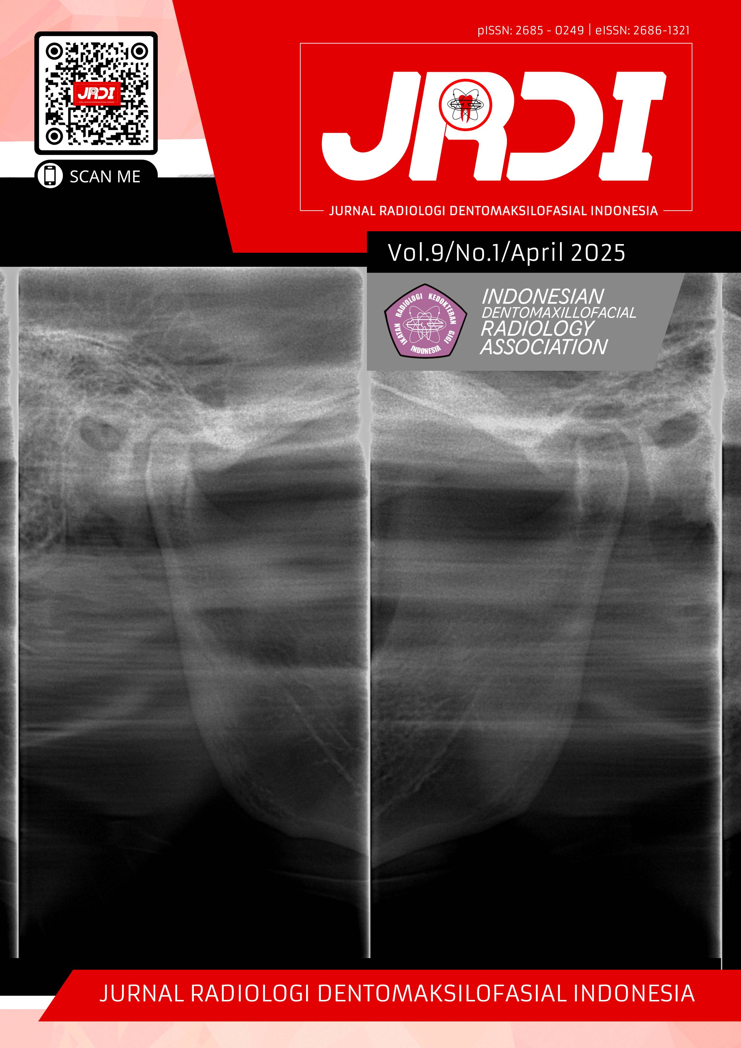
Radiographic finding of sunray appearance as a sign of malignant mandibular lesion: a case report
Abstract
Objectives: To report the “sunray” appearance on panoramic radiography as a sign of malignancy lesions of the mandible.Case Report: A 40 year old female patient came to the Hasanuddin University Dental Hospital with the main complaint of facial swelling which causes an asymmetrical appearance and hard consistency on palpation. Mucosa around the second right premolar to the third right molar is reddish with an irregular border. The patient was referred to the radiology department for panoramic radiography and MRI. The panoramic radiograph revealed a mixed radiolucent-radiopaque lesion in tooth 35 involving ramus to the coronoid process. PDL space was irregular widening at 36, 37, and 38. The "sunray” appearance was seen from the ramus extending to the coronoid process. On the MRI, a mass on the submandibular gland pushed and narrowed the sublingual, parapharyngeal, and masticator space, destroying the mandible on the left side. These radiologic findings strongly suggest a malignancy involving the jawbone.
Conclusion: The findings of a mandibular malignancy in the form of a “sunray” appearance on panoramic radiography need to be confirmed with an MRI examination to determine the consistency and extent of the lesion to the surrounding tissue. A comprehensive examination is necessary to properly diagnose mandibular malignant lesions so that the most suitable treatment plan can be determined.
References
Epsilawati L, Firman R, Pramanik F, Ambarlita Y. Gambaran radiograf dari lesi keganasan maksilofasial: (literature review). Makassar Dent J. 2018;7(2):88–94.
White SC, Pharoah MJ. Oral Radiology: Principles and Interpretation. 7th ed. St. Louis: Elsevier Health Sciences; 2014.
Athar M, Chauhan S, Tripathi R, Kala S, Avasthi S, Jangra V, et al. Malignant mandibular tumors: two case reports of rare mandibular tumors in a single institution. Arch Med Biomed Res. 2014;1(1):22–9.
Nurrachman AS, Pramanik F, Azhari A, Epsilawati L. Gambaran border dan periosteal reaction lesi rahang pada radiograf. J Radiol Dentomaksilofasial Indones. 2020;4(1):31.
Rego Costa F, Esteves C, Bacelar MT. Benign mandibular lesions: a pictorial review. Acta Radiol Port. 2016;28:45–52.
Widyaningrum R, Faisal A, Mitrayana M, Mudjosemedi M, Agustina D. Oral cancer imaging: the principles of interpretation on dental radiograph, CT, CBCT, MRI, and USG. Maj Kedokt Gigi Indones. 2018;4(1):1–14.
Maia Ferreira Alencar CH, D'Ippolito G, Carneiro A, da Fonseca LMB, de Almeida M, de Melo ASA. Periosteum: an imaging review. Eur J Radiol Open. 2020;7:100249.
Kakarla SK, Suneetha P, Prasad SS, Reddy MR. Imaging spectrum of periosteal lesions: pictorial essay. J Med Sci Res. 2020;8(3):121–38.
Aicha Z, Souha BY, Kaoutharn S, Wafa H, Chedly B. Jaw malignancies: signs that should alert the dentist. Tunis Med. 2011;89(7):580–4.
Lestari S. Analisis informasi fisis radiograf panoramik digital untuk deteksi tumor jinak pada rahang. [Tesis]. [Indonesia]: Universitas Gadjah Mada; 2019.
Mortazavi H, Safi Y, Rahmani S, Rezaiefar K. Oral hard tissue lesions: a radiographic diagnostic decision tree. Open Access Maced J Med Sci. 2020;8(F):180–6.
Samraj L, Kaliamoorthy S, Venkatapathy R, Oza N. Osteosarcoma of the mandible: a case report with an early radiographic manifestation. Imaging Sci Dent. 2014;44(1):85–8.
Loubna A, Bouchra T. Osteosarcoma of the jaws: a case report. Int J Surg Case Rep. 2022;93:106993.
Rančić N, Stojanović M, Petrović B, Stojanović-Radić Z, Jovanović M, Rančić Z. Radiological characteristics of periosteal reactions. Acta Med Medianae. 2020;59(4):116–24.
de Sá Neto JL, Simão MN, Crema MD, Engel EE, Nogueira-Barbosa MH. Desempenho diagnóstico da ressonância magnética na avaliação de reações periosteais em sarcomas ósseos utilizando a radiografia convencional como padrão de referência. Radiol Bras. 2017;50(3):176–81.
Basaran B, Doruk C, Yilmaz E, Sunnetcioglu E, Bilgic B. Intraosseous mucoepidermoid carcinoma of the jaw: report of three cases. Turk Arch Otorhinolaryngol. 2018;56(1):42–6.
Singh H, Yadav AK, Chand S, Singh A, Shukla B. Central mucoepidermoid carcinoma: case report with review of literature. Natl J Maxillofac Surg. 2019;10(1):109–13.
De Freitas GB, de Souza LL, de Souza FJ, de Almeida OP, Lopes MA. Intraosseous mucoepidermoid carcinoma in the mandible. Case Rep Dent. 2018;2018:1–4.
Khataniar H, Senthil S, Deep SS, Ramesh R, Y K I. Intraosseous mucoepidermoid carcinoma of the anterior mandible: a case report. Cureus. 2022;14(5):e25036.
Published
2025-05-31
How to Cite
ALFINA, Risca et al.
Radiographic finding of sunray appearance as a sign of malignant mandibular lesion: a case report.
Jurnal Radiologi Dentomaksilofasial Indonesia (JRDI), [S.l.], v. 9, n. 1, p. 29-32, may 2025.
ISSN 2686-1321.
Available at: <http://jurnal.pdgi.or.id/index.php/jrdi/article/view/1249>. Date accessed: 09 feb. 2026.
doi: https://doi.org/10.32793/jrdi.v9i1.1249.
Section
Case Report

This work is licensed under a Creative Commons Attribution-NonCommercial-NoDerivatives 4.0 International License.


