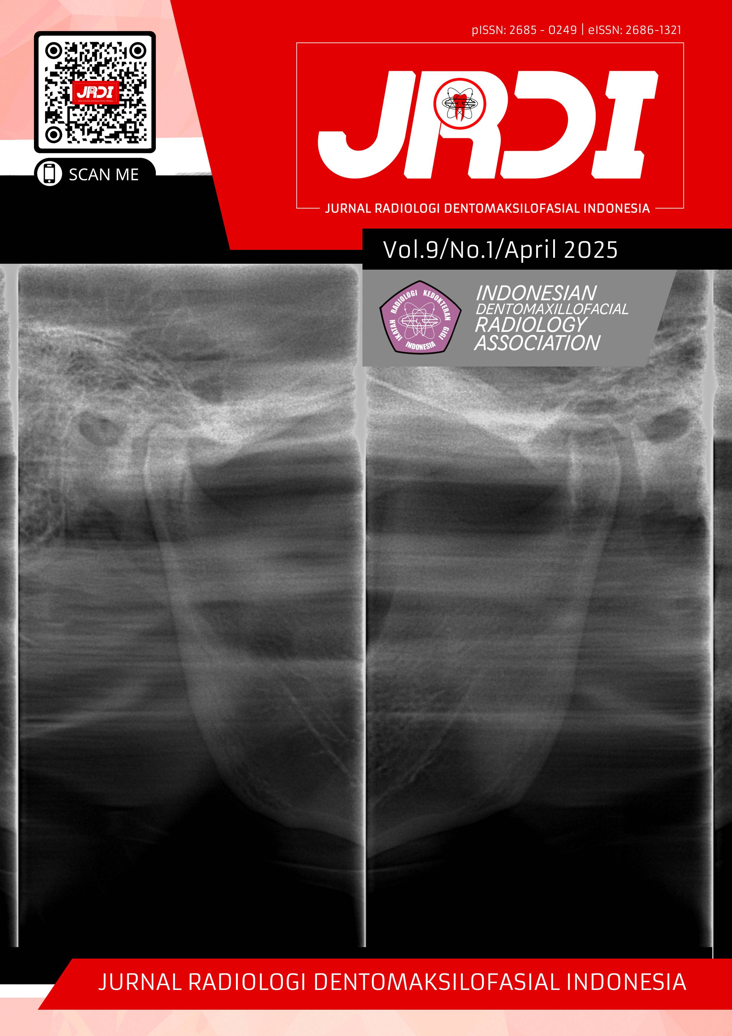
Mandibular osteomyelitis on panoramic radiographs: a case series
Abstract
Objectives: This series of cases aims to see an extension of a lesion using panoramic radiographs to help establish a diagnosis in three cases in the mandible. Osteomyelitis occurs more often in the mandible than in the maxilla because the maxilla has a better blood supply than the mandible, with relatively thinner cortical and fewer medullary cavities.Case Report: Three female patients presented with nearly identical complaints, including frequent pus discharge, unpleasant odor, and swelling. Only two of the patients had a history of tooth extraction. All three were referred to the university dental hospital (RSGM) for further management. Panoramic radiographs revealed similar findings among the three cases, including mixed radiopaque-radiolucent images with irregular shapes and diffuse borders. In two patients, sequestra were visible in the right and left corpus regions of the mandible. In contrast, the third patient showed a slightly different presentation: a well-defined, irregular radiopaque mass protruding from the top of the alveolar bone, localized at the crest of the ridge. One of the patients had a history of systemic diseases, specifically hypertension which was under control.
Conclusion: In these three cases, a panoramic X-ray examination was the only support for identifying the characteristics of lesion expansion and was considered sufficient as a reference for patient management. However, a definitive diagnosis still requires a histopathological examination.
References
Jung GW, Moon SY, Oh JS, Choi HI, You JS. Acute osteomyelitis of the mandible by extended-spectrum β-lactamase-producing Klebsiella pneumoniae: a case report. J Oral Med Pain. 2021;46(3):88–92.
Baltensperger M, Eyrich G. Osteomyelitis of the jaws: definition and classification. In: Osteomyelitis of the Jaws. Berlin: Springer; 2009. p.5–56.
Sudirman PL. Osteomielitis maksila: laporan kasus dari tiga kasus. Denpasar: Fakultas Kedokteran Universitas Udayana; 2016.
Simakerti G. Gambaran radiologi osteomielitis. Bandung: Departemen Radiologi Fakultas Kedokteran Universitas Padjadjaran RS Dr. Hasan Sadikin; 2019. p. 9–10.
Uçkay I, Lew D, Suva D, Vaudaux P, Kronig I. Acute and chronic osteomyelitis. Geneva: Geneva University Hospitals and Faculty of Medicine; 2015.
van der Meent M, Pichardo SEC, Dijkstra NMA, van Merkesteyn JPR. Outcome of different treatments for chronic diffuse sclerosing osteomyelitis of the mandible: a systematic review. Br J Oral Maxillofac Surg. 2020;58(4):385–90.
Barajas-Pérez VH, Recendez-Santillan NJ, Vega-Memíje ME, García-Calderón AG, Cuevas-González JC. Chronic suppurative osteomyelitis of the mandible treated with antibiotics complemented with surgical treatment: a case report. Int J Odontostomat. 2017;11(3):261–5.
Ayekinam KAO, Saliha C, Wafaa EW. Chronic suppurative osteomyelitis in extraction molar mandible complications: a case report. JMSR. 2017;4(2):457–60.
Dym H, Zeidan J. Microbiology of acute and chronic osteomyelitis and antibiotic treatment. Dent Clin North Am. 2017;61(2):271–82.
Gudmundsson T, Torkov P, Thygesen TH. Diagnosis and treatment of osteomyelitis of the jaw: a systematic review. J Dent Oral Disord. 2017;3(3).
Dasam S, Ramesh T, Kishore M, Manyam R. Osteomyelitis of maxilla: an unusual case. J Indian Acad Oral Med Radiol. 2020.
Park MS, Eo MY, Myoung H, Kim SM, Lee JH. Early diagnosis of jaw osteomyelitis by easy digitalized panoramic analysis. Maxillofac Plast Reconstr Surg. 2019;41:6.
Lestari S. Analisis informasi fisis radiograf panoramik digital untuk deteksi tumor jinak pada rahang. J Teknol Inf. 2015;X(30):1–6.
Gupta D. Role of maxillofacial radiology and imaging in the diagnosis and treatment of osteomyelitis of the jaws. M.M. College of Dental Sciences and Research; 2015 Jul 16.
Putri NPSS, Yunus B. Penggunaan teknik radiografi konvensional dan digital pada perawatan endodontik: tinjauan pustaka. Cakradonya Dent J. 2021;13(2):97–105.
Silva BSF, Bueno MR, Yamamoto-Silva FP, Gomez RS, Peter OA, Estrela C. Differential diagnosis and clinical management of periapical radiopaque/hyperdense jaw lesions. J Appl Oral Sci. 2017 May 31.
Rochmah YS. Osteomyelitis kronis mandibula pasca ekstraksi gigi disertai Bell’s palsy. ODONTO Dent J. 2019;6(1):1–6.
Buch K, Thuesen ACB, Brons C, Schwarz P. Chronic non-bacterial osteomyelitis: a review. Calcif Tissue Int. 2019;104(5):544–53.
Shin JW, Kim JE, Huh KH, Yi WJ, Heo MS, Lee SS, Choi SC. Clinical and panoramic radiographic features of osteomyelitis of the jaw: a comparison between antiresorptive medication-related and unrelated conditions. Imaging Sci Dent. 2019;49(4):287–94.
van der Meent MM, Dijkstra NM, Glas MJM, Pichardo SEC, van Merkesteyn JPR. Bisphosphonate therapy in chronic diffuse sclerosing osteomyelitis/tendoperiostitis of the mandible: retrospective case series. J Craniomaxillofac Surg. 2022;50(5):433–40.
Zimmermann C, Stuepp RT, Silva Rath IBD, Grando LJ, Daniel FI, Meurer MI. Diagnostic challenge and clinical management of juvenile mandibular chronic osteomyelitis. Head Neck Pathol. 2019.
Simanjuntak HF, Sylvyana M, Fathurachman. Osteomyelitis kronis supuratif mandibula sebagai komplikasi sekunder impaksi gigi molar tiga. Maj Kedokt Gigi Komunitas. 2016;2(1):13–8.
Chattopadhyay PK, Nagori SA, Menon RP, Balasundaram T. Osteomyelitis of the mandibular condyle: a report of 2 cases with review of the literature. J Oral Maxillofac Surg. 2016;74(12):2569.e1–6.
Mallya SM, Lam EWN. White and Pharoah’s Oral Radiology: Principles and Interpretation. 8th ed. St. Louis: Elsevier; 2019. p.906.
Published
2025-06-09
How to Cite
THALA, Ifa Ariefah Hs; WULANSARI, Dwi Putri; RASUL, Muhammad Irfan.
Mandibular osteomyelitis on panoramic radiographs: a case series.
Jurnal Radiologi Dentomaksilofasial Indonesia (JRDI), [S.l.], v. 9, n. 1, p. 47-54, june 2025.
ISSN 2686-1321.
Available at: <http://jurnal.pdgi.or.id/index.php/jrdi/article/view/1264>. Date accessed: 09 feb. 2026.
doi: https://doi.org/10.32793/jrdi.v9i1.1264.
Section
Case Report

This work is licensed under a Creative Commons Attribution-NonCommercial-NoDerivatives 4.0 International License.


