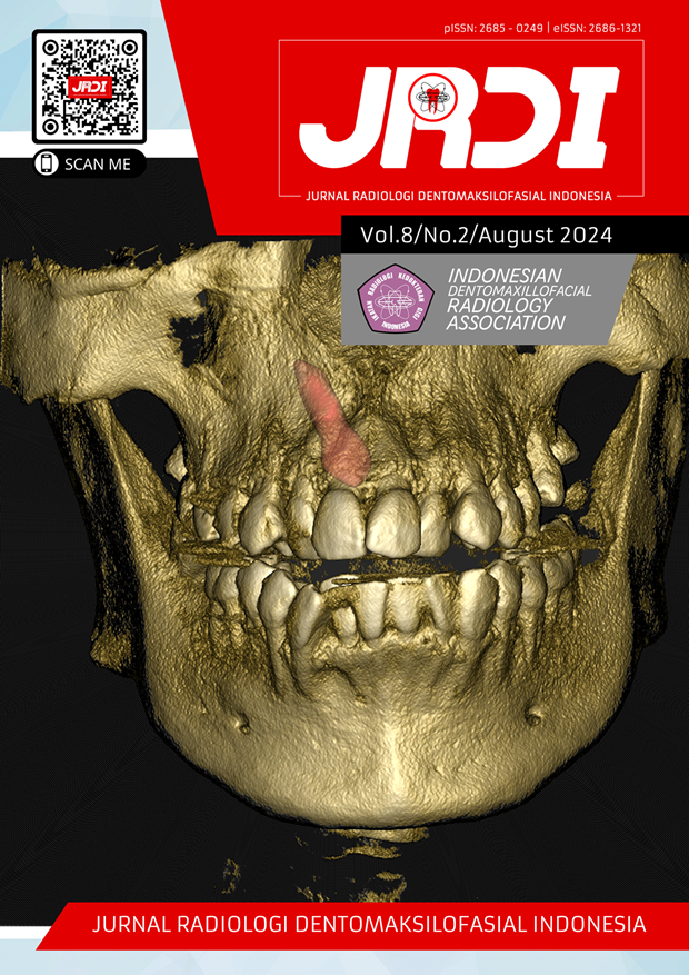
Ameloblastoma radiographic imaging on 3D CBCT: a literature review
Abstract
Objectives: This review article is aimed to provide an overview of 3D CBCT in determining the diagnosis of ameloblastoma.Review: This study is a literature review consisting of English articles about the ameloblastoma radiographic imaging on 3D CBCT, published 2013-2023.The article search databases used were Google scholar, Ebsco, Pubmed .The total search results for articles based on keywords obtained were 552 articles, and only 9 articles were include. Ameloblastoma is a persistent and locally invasive tumor; with aggressive but docile growth characteristics. Ameloblastoma is generally associated with impacted teeth, so it requires a more detailed radiographic examination. Computed cone-beam tomography (CBCT) is a more advantageous imaging system with a lower radiation dose and smaller area requirements. 3D CBCT is a radiographic examination with a high modality, so it is very important in helping to establish a diagnosis, especially for cases that show radiographic differences. Ameloblastoma is divided into several types based on the radiological picture. Odontogenic Keratosis Cysts and ameloblastoma may exhibit similar radiographic features, which make diagnosis difficult.
Conclusion: 3D CBCT examination is helpful in diagnosing and validating the treatment of ameloblastoma.
References
Tenorio JR, Velasco SK, Nunes SD, Cavalcanti MGP. Radiographic imaging pattern of ossifying fibroma mimicking ameloblastoma: a case report. Clin Lab Res Den 2019:1-5.
Riachi F, Khairallah CM, Ghosn N, Berberi AN. Cyst volume changes measured with a 3D reconstruction after decompression of a mandibular dentigerous cyst with an impacted third molar. Clin Pract. 2019;9(1):1132.
Kişa HC , Coşgunarslan A , Canger EM, Etöz M , Deniz K. Management of Ameloblastoma with Different Imaging Modalities. Turkiye Klinikleri J Case Rep. 2020;28(3):145-55.
Madiyal A, Babu SG, Castelino R, Ajila V, Bhat S, Achalli S, Madi M, Rao K. Use of cone beam computed tomography in the diagnosis and treatment planning of follicular ameloblastoma: A case report with review of literature. J Turgut Ozal Med Cent . 2017;24(3):333-7.
Siregar M, Sitam S, Lita YA, Hadikrishna I. Analysis Of Dentigerous Cyst, Ameloblastoma, And Odontogenic Keratocyst Panoramic Radiograph And CBCT: A Scoping Review. Odonto Dental Journal. 2022;9(1):115-30.
Luo J, You M, Zheng G, Xu L. Cone beam computed tomography signs of desmoplastic ameloblastoma: review of 7 cases. Oral Surg Oral Med Oral Pathol Oral Radiol. 2014;118(4):e126-33.
Sharma A, Nair S, Shah A; Kumar. Amelobastoma of the Mandible in a Pediatric Patient : A Case Report with Review of Literature. Clove Dental Journal Of Clinical Dentistry. 2015;1(1):34-40.
Liaquat A, Baig MA, Mehmood A, Saeed T. Solid Ameloblastoma: A Case Report. Archives of Surgical Research. 2020;1(4):41-4.
Rezaei MM, Bagherpour A, Mahmoudi P. Ameloblastoma ex calcifying odontogenic cyst in the mandible: report of a rare case. Cumhuriyet Dent J. 2014;17:84-91.
Kang BC, Lee JS, Yoon SJ, Kim Y. Ameloblastoma with dystrophic calcification: A case report with 3-dimensional cone-beam computed tomographic images of calcification. Imaging Sci Dent. 2020;50(4):373-6.
Krishnamoorthy B, Sharma H, Suma GN, Kukreja R. Report of Unicystic Ameloblastoma of Mandible in a Young Adult – A Radiological Perspective. Int J Oral Health Med Res. 2015;2:63-6.
Kitisubkanchana J, Reduwan NH, Poomsawat S, Pornprasertsuk-Damrongsri S, Wongchuensoontorn C. Odontogenic keratocyst and ameloblastoma: radiographic evaluation. Oral Radiol. 2021;37(1):55-65.
Kumar A, Venkatesh E, Srikanth MDM, Fatima N. An Unusual Case Report of a Unicystic Ameloblastoma in the Body of Mandible Masquerading as Radicular Cyst and its Evaluation with Cone Beam Computed Tomography. Universal Research Journal of Dentistry. 2013;3(1):41-3.
Pagare J, Johaley S. Cone Beam Computed Tomography- A Boon in Diagnosis of Expansile Follicular Ameloblastoma of Mandible - A Case Report. International Journal of Research and Review. 2019;6(8):198-202.
Nagarajan N, Sadaksharam J. Current Concepts in Imaging and Management of Ameloblastoma. Medical Reports and Case Studies. 2021;06(2):001-2.
Alves DBM, Tuji FM, Alves FA, Rocha AC, Santos-Silva ARD, Vargas PA, Lopes MA. Evaluation of mandibular odontogenic keratocyst and ameloblastoma by panoramic radiograph and computed tomography. Dentomaxillofac Radiol. 2018;47(7):20170288.
Mathew J, Akhil S, Thomas J, Ali F. Ravaged mandibular ramus: Two rare case presentations of unicystic ameloblastoma with a view on management. Journal of Oral Research and Review. 2022;14(2):145-9.
Meng Y, Zhao YN, Zhang YQ, Liu DG, Gao Y. Three-dimensional radiographic features of ameloblastoma and cystic lesions in the maxilla. Dentomaxillofac Radiol. 2019;48(6):20190066.
Candido IR, da Silva CSV, Dos Santos Garcia E, da Silva ALF, Fernandes Poleti TMF, Gialain IO, Borba AM. The Usefulness of a Facial Digital Biobank for Ameloblastoma Resection and Fracture Fixation - A Case Report. Ann Maxillofac Surg. 2021;11(2):325-8.
Brown AA, Scarfe WC, Scheetz JP, Silveira AM, Farman AG. Linear accuracy of cone beam CT derived 3D images. Angle Orthod. 2009;79(1):150-7.
Thengumpallil S, Smith K, Monnin P, Bourhis J, Bochud F, Moeckli R. Difference in performance between 3D and 4D CBCT for lung imaging: a dose and image quality analysis. J Appl Clin Med Phys. 2016;17(6):97-106.
Raj Subash C, Das Surya N, Patnaik K, Panda Subhasree M, Praharaj K. Peripheral Ameloblastoma With Interdental Bone Loss: A Rare Case Report. International Journal of Current Research. 2017;9(07);54966-8.
Guo T, Zhang C, Zhou J. Unicystic ameloblastoma in a 9-year-old child treated with a combination of conservative surgery and orthodontic treatment: A case report. Clin Case Rep. 2022;10(1):e05241.
Published
2024-08-20
How to Cite
UKE, Waode Anita Wulanduri; YUNUS, Barunawaty.
Ameloblastoma radiographic imaging on 3D CBCT: a literature review.
Jurnal Radiologi Dentomaksilofasial Indonesia (JRDI), [S.l.], v. 8, n. 2, p. 89-96, aug. 2024.
ISSN 2686-1321.
Available at: <http://jurnal.pdgi.or.id/index.php/jrdi/article/view/1273>. Date accessed: 25 feb. 2026.
doi: https://doi.org/10.32793/jrdi.v8i2.1273.
Section
Review Article

This work is licensed under a Creative Commons Attribution-NonCommercial-NoDerivatives 4.0 International License.


