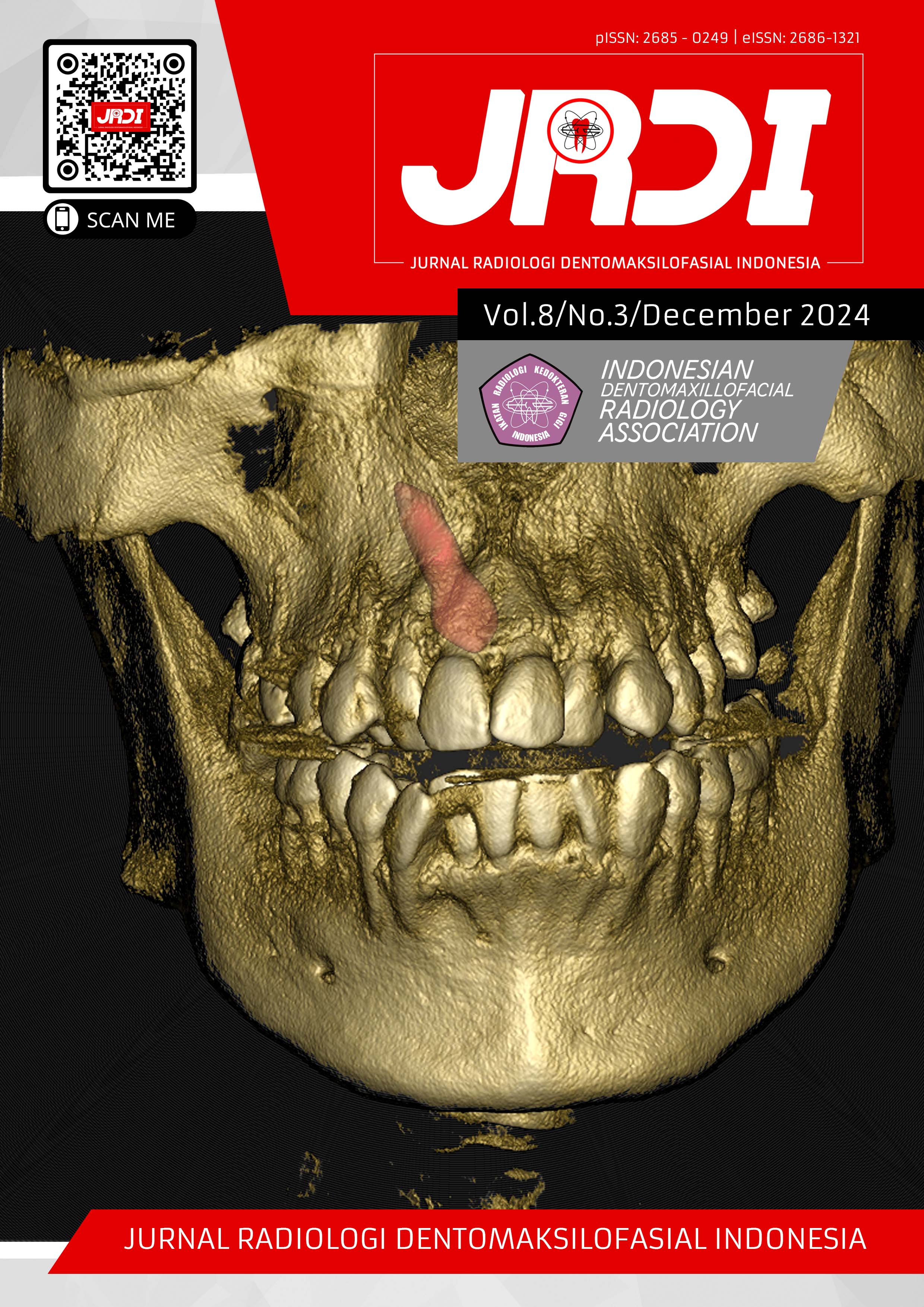
The role of radiographic imaging and finite element analysis in evaluating occlusal loads and stress distribution in the periodontal ligament
Abstract
Objectives: Biomechanical behavior analysis of the periodontal ligament (PDL) under various loading conditions is essential for understanding the impact of occlusal force distribution. A comprehensive understanding of this aspect is fundamental, and radiographic examination is a crucial modality for evaluating periodontal health. This review aims to illustrate the role of radiographic examination in influencing dental prognosis through the use of Finite Element Analysis (FEA) to assess occlusal load and stress distribution in PDLs.Review: Radiographic imaging techniques are critical for assessing the extent of occlusal trauma and its impact on the periodontal ligament and surrounding structures. Modalities such as conventional radiography, cone-beam computed tomography (CBCT), and micro-computed tomography (micro-CT) are commonly used to evaluate occlusal load. Studies have demonstrated that a balanced occlusal scheme results in a more uniform stress distribution, while an unbalanced scheme leads to localized stress concentrations, increasing the risk of periodontal damage. FEA has emerged as a powerful tool for simulated and visualizing stress patterns in the PDL and quantitatively calculating stresses and deformations in the periodontium. Technological advances in imaging, when applied in conjunction with finite element computational techniques, have shown that oblique loading results in higher stress concentrations compared to vertical loading, particularly in the PDL of mandibular first molars. These higher stresses, often observed in the cervical and apical regions, highlight the potential for more significant PDL damage, making it useful for evaluating bone loss and PDL integrity. for eligibility and completeness of journals.
Conclusion: Integration of advance radiographic imaging with FEA has significantly enhanced the understanding of occlusal load and stress distribution in the periodontal ligament. This advancement has propelled the field of periodontal biomechanics, offering very valuable insights into PDL’s biomechanical behavior as it responds to varying occlusal loads, to optimize outcomes in periodontal and orthodontic care.
References
Vandana K, Muneer S. Effect of Different Occlusal Loads on Periodontium: A Three-dimensional Finite Element Analysis. CODS Journal of Dentistry. 2016;8(2):78-90.
Carranza M, Newman G, Takei H, Perry R, Klokkevold A, Carranza F. Clinical Periodontology.; 2015.
Nanci Antonio. Ten Cate’s Oral Histology : Development, Structure, and Function. Elsevier; 2018.
Poiate IAVP, de Vasconcellos AB, de Santana RB, Poiate E. Three‐Dimensional Stress Distribution in the Human Periodontal Ligament in Masticatory, Parafunctional, and Trauma Loads: Finite Element Analysis. J Periodontol. 2009;80(11):1859-67.
Lang NP, Bartold PM. Periodontal health. J Periodontol. 2018;89:S9-S16.
Reddy RT, Vandana KL. Effect of hyperfunctional occlusal loads on periodontium: A three-dimensional finite element analysis. J Indian Soc Periodontol. 2018;22(5):395.
Mallya SM;, Lam EWN. White and Pharoah’s Oral Radiology, Principle and Interpretation.; 2014.
Vijay G, Raghavan V. Radiology in Periodontics. Journal of Indian Academy of Oral Medicine and Radiology. 2013;25:24-9.
Vandana K, Muneer S. Effect of Different Occlusal Loads on Periodontium: A Three-dimensional Finite Element Analysis. CODS Journal of Dentistry. 2016;8(2):78-90.
Reddy RT, Vandana KL. Effect of hyperfunctional occlusal loads on periodontium: A three-dimensional finite element analysis. J Indian Soc Periodontol. 2018;22(5):395-400.
Atif M, Tewari N, Reshikesh M, Chanda A, Mathur VP, Morankar R. Methods and applications of finite element analysis in dental trauma research: A scoping review. Dental Traumatology. Published online 2024.
Mortazavi H, Baharvand M. Review of common conditions associated with periodontal ligament widening. Imaging Sci Dent. 2016;46(4):229-37.
Ortún-Terrazas J, Cegoñino J, Pérez del Palomar A. In silico study of cuspid’ periodontal ligament damage under parafunctional and traumatic conditions of whole-mouth occlusions. A patient-specific evaluation. Comput Methods Programs Biomed. 2020;184.
Robo I, Heta S. Radiographic Signs of Trauma from Occlusion at the Level of the Periodontal Ligament and Articulation Structures. Journal of Case Reports and Medical Images. 2020;3(1):1049.
Shahidi S, Zamiri B, Abolvardi M, Akhlaghian M, Paknahad M. Comparison of Dental Panoramic Radiography and CBCT for Measuring Vertical Bone Height in Different Horizontal Locations of Posterior Mandibular Alveolar Process. J Dent (Shiraz). 2018;19(2):83-91.
Corbet EF, Ho DKL, Lai SML. Radiographs in periodontal disease diagnosis and management. Aust Dent J. 2009;54:S27-S43.
Campioni I, Pecci R, Bedini R. Ten years of micro-CT in dentistry and maxillofacial surgery: A literature overview. Applied Sciences (Switzerland). 2020;10(12).
Li Y, Zhan Q, Bao M, Yi J, Li Y. Biomechanical and biological responses of periodontium in orthodontic tooth movement: up-date in a new decade. Int J Oral Sci. 2021;13(1).
Shukla S, Chug A, Afrashtehfar KI. Role of cone beam computed tomography in diagnosis and treatment planning in dentistry: An update. J Int Soc Prev Community Dent. 2017;7:S125-36.
Acar B. Use of cone beam computed tomography in periodontology. World J Radiol. 2014;6(5):139.
Narayana Prasad P, Sharma T, Aggarwal A, Rana T, Dabla N, Rawat N. A Three Dimensional Finite Element Analysis For Stress In Periodontal Tissues Of Maxillary 1st Molar By Intrusive Forces Using TADS-FEA Study A Three Dimensional Finite Element Analysis For Stress In Periodontal Tissues Of Maxillary 1 ST Molar By Intrusive Forces Using TADS-FEA Study. J Clin Den Res Edu. 2013;2(5):6-12.
Zhang H, Cui JW, Lu XL, Wang MQ. Finite element analysis on tooth and periodontal stress under simulated occlusal loads. J Oral Rehabil. 2017;44(7):526-36.
Tsai MT, He RT, Huang HL, Tu MG, Hsu JT. Effect of Scanning Resolution on the Prediction of Trabecular Bone Microarchitectures Using Dental Cone Beam Computed Tomography. Diagnostics journal. 2020;10(368):1-7.
Flugge T, Gross C, Ludwig U, et al. Dental MRI-only a future vision or standard of care? A literature review on current indications and applications of MRI in dentistry. Dentomaxillofacial Radiology. 2023;52(4).
Dewake N, Miki M, Ishioka Y, Nakamura S, Taguchi A, Yoshinari N. Association between clinical manifestations of occlusal trauma and magnetic resonance imaging findings of periodontal ligament space. Dentomaxillofacial Radiology. 2023;52(8).
Boas FE, Fleischmann D. CT artifacts: Causes and reduction techniques. Imaging Med. 2012;4(2):229-40.
Reinhardt RA, Pao YC, Krejci RF. Periodontal Ligament Stresses in the Initiation of Occlusal Traumatism. Vol 19.; 1984.
Geramy A, Faghihi S. Secondary trauma from occlusion: Three-dimensional analysis using the finite element method. Quintessence Int (Berl). 2004;35(10):835-43.
Oyama K, Motoyoshi M, Hirabayashi M, Hosoi K, Shimizu N. Effects of root morphology on stress distribution at the root apex. Eur J Orthod. 2007;29(2):113-7.
Benazzi S, Nguyen HN, Kullmer O, Kupczik K. Dynamic modelling of tooth deformation using occlusal kinematics and finite element analysis. PLoS One. 2016;11(3).
Liu H, Jiang H, Wang Y. The biological effects of occlusal trauma on the stomatognathic system - a focus on animal studies. J Oral Rehabil. 2013;40(2):130-8.
Chen YC, Tsai HH. Use of 3D finite element models to analyze the influence of alveolar bone height on tooth mobility and stress distribution. J Dent Sci. 2011;6(2):90-4.
Pini M, Zysset P, Botsis J, Contro R. Tensile and compressive behaviour of the bovine periodontal ligament. J Biomech. 2004;37(1):111-9.
Murakami K, Yamamoto K, Sugiura T, et al. Effect of clenching on biomechanical response of human mandible and temporomandibular joint to traumatic force analyzed by finite element method. Med Oral Patol Oral Cir Bucal. 2013;18(3).
Baghani Z, Soheilifard R, Bayat S. How Does the First Molar Root Location Affect the Critical Stress Pattern in the Periodontium? A Finite Element Analysis. Journal of Dentistry (Iran). 2023;24(2):182-93.
Ahmić Vuković A, Jakupović S, Zukić S, Bajsman A, Gavranović Glamoč A, Šečić S. Occlusal Stress Distribution on the Mandibular First Premolar - FEM Analysis. Acta Med Acad. 2019;48(3):255-261.
Lee H, Kim M, Chun YS. Comparison of occlusal contact areas of class I and class II molar relationships at finishing using three-dimensional digital models. Korean J Orthod. 2015;45(3):113-20.
Natali AN, Pavan PG, Scarpa C. Numerical analysis of tooth mobility: Formulation of a non-linear constitutive law for the periodontal ligament. Dental Materials. 2004;20(7):623-9.
Moga RA, Olteanu CD, Botez MD, Buru SM. Assessment of the Orthodontic External Resorption in Periodontal Breakdown—A Finite Elements Analysis (Part I). Healthcare (Switzerland). 2023;11(10).
Published
2025-01-01
How to Cite
NAINGGOLAN, Lidya Irani et al.
The role of radiographic imaging and finite element analysis in evaluating occlusal loads and stress distribution in the periodontal ligament.
Jurnal Radiologi Dentomaksilofasial Indonesia (JRDI), [S.l.], v. 8, n. 3, p. 133-140, jan. 2025.
ISSN 2686-1321.
Available at: <http://jurnal.pdgi.or.id/index.php/jrdi/article/view/1299>. Date accessed: 25 feb. 2026.
doi: https://doi.org/10.32793/jrdi.v8i3.1299.
Section
Review Article

This work is licensed under a Creative Commons Attribution-NonCommercial-NoDerivatives 4.0 International License.


