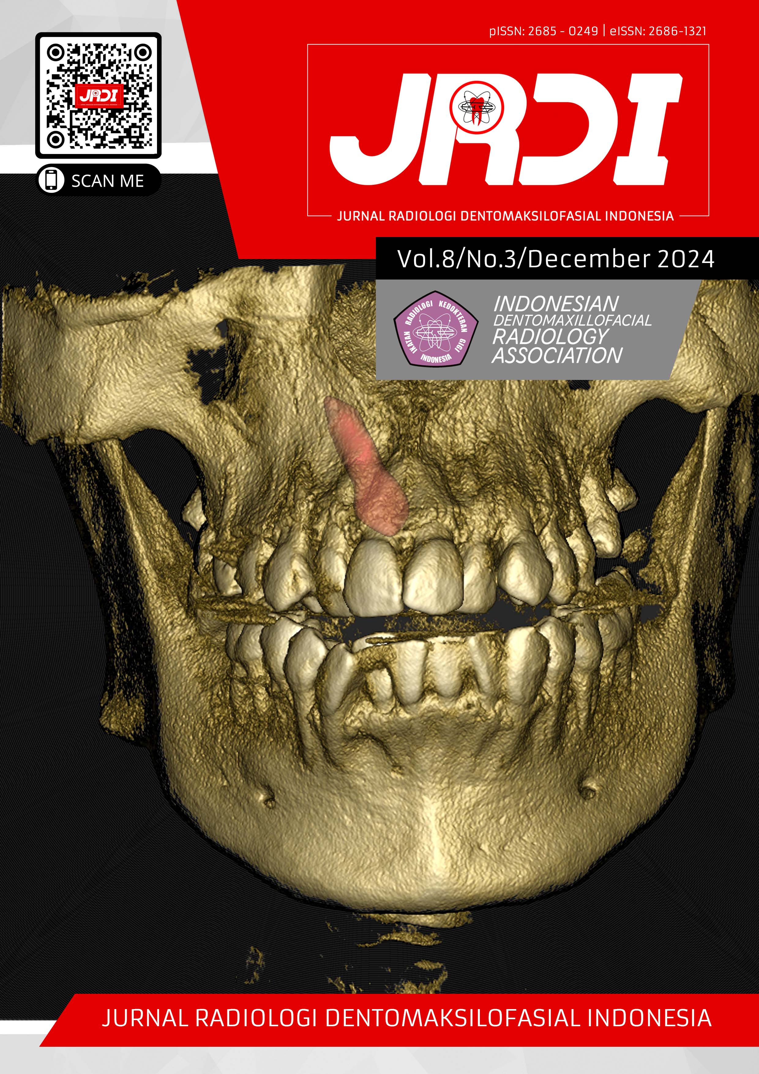
Description of the shape and position of the condyles in Kennedy classification class I, II, III, and IV patients through panoramic radiography
(At RSUD Ulin and RSGM Gusti Hasan Aman Banjarmasin from January 2018 - January 2024)
Abstract
Objectives: Tooth loss occurs when the tooth detaches from the socket. Cases of partial tooth loss can cause differences in the shape and position of the condyles. This study aimed to know the description of the frequency distribution of normal and abnormal condyle shapes and positions in Kennedy classification case patients class I, II, III, IV.Materials and Methods: This research used a cross-sectional descriptive approach. The sample used secondary data from 120 digital panoramic radiographic photos of patients aged 30-70 from January 2018 to January 2024 at Ulin Hospital and Gusti Hasan Aman Hospital Banjarmasin.
Results: Based on the research results at RSUD Ulin and RSGM Gusti Hasan Aman Banjarmasin, the round shape was the most common condyle shape found in patients with Kennedy classification, with most condyle positions pointing to the anterior. The change in the shape and position of the condyle becomes pathological due to the long-term loss of part of the tooth.
Conclusion: The frequency distribution of the shape and position of the condyle of patients with Kennedy classification class I, II, III, IV was the round shape as the most common condyle shape experienced by patients which is one of the normal condyles shapes, and an abnormal position of TMJ condition pointing anteriorly.
References
WHO. Oral Health. World Health Organization. 2023. Available from: https://www.who.int/news-room/fact-sheets/detail/oral-health
Kementrian Kesehatan RI. RISKESDAS 2018. Jakarta: Badan Penelitian dan Pengembangan Kesehatan; 2018. P.184–5.
Kementrian Kesehatan RI. Laporan provinsi Kalimantan Selatan RISKESDAS 2018. Badan Litbang Kesehatan; 2019. p.133–4.
Siagian K. Kehilangan sebagian gigi pada rongga mulut. Jurnal e-Clinic (eCl). 2016 Jun;4(1):1–6.
Lai S, Damayanti L, Wulansari D. Gangguan sendi temporomandibular akibat ruang edentulous pada usia dewasa muda. Padjadjaran Journal of Dental Researchers and Students. 2023 Mar 3;7(1):13–7.
Ramadhan R, Pramanik F, Epsilawati L. Radiograf panoramik digital bentuk kepala kondilus pada pasien kliking dan tidak kliking. Padjadjaran Journal of Dental Researchers and Students. 2019 Nov 9;3(2):134–40.
Puspitasari G, Damayanti L, Kusumadewi AN. Pola kehilangan gigi berdasarkan klasifikasi Kennedy serta penyebab utama kehilangan gigi pada rahang atas atau rahang bawah usia dewasa muda. Jurnal Kedokteran Gigi Universitas Padjajaran. 2022 Dec 30;34(3):216–23.
Rahmayani L, Andriany P. Distribusi frekuensi kehilangan gigi berdasarkan klasifikasi Kennedy ditinjau dari tingkat pendapatan masyarakat Kelurahan Peuniti Banda Aceh. ODONTO Dental Journal. 2015 Jul;2(1):8–13.
Anjani K, Nurrachman A, Rahman F, Firman R, dkk. Bentuk dan posisi kondilus sebagai marker pada temporomandibular disorder (TMD) melalui radiografi panoramik. Jurnal Radiologi Dentomaksilofasial Indonesia (JRDI). 2020 Dec 30;4(3):91.
Li D, Leung Y. Temporomandibular disorders: Current concepts and controversies in diagnosis and management. Diagnostics. 2021 Mar 1;11(3):1–15.
Shofi N, Sukmana I. Deskripsi kasus temporomandibular disorder pada pasien di RSUD Ulin Banjarmasin bulan Juni – Agustus 2013 tinjauan berdasarkan jenis kelamin, etiologi, dan klasifikasi. Dentino. 2014 Mar;2(1):70–3.
Darmawan F, Indah M, Irianty H. Faktor-faktor yang berhubungan dengan tindakan pengelolaan sampah medis benda tajam di Rumah sakit Ulin Banjarmasin tahun 2020. [Banjarmasin]: Universitas Islam Kalimantan Selatan; 2020.
RSGM Gusti Hasan Aman – Selalu sedia melayani anda [cited 2024 Jul 20]. Available from: https://rsgm.kalselprov.go.id/
Hikmawati F. Metodologi penelitian. 1st ed. Vol. 4. Depok: PT RajaGrafindo Persada; 2020. 13–120 p.
Abdullah M. Metodologi penelitian kuantitatif. 1st ed. Istiadi Agung, Igbal, editors. Vol. 1. Yogyakarta: Aswaja Pressindo; 2015. 1–409 p.
Syapitri H, Amila, Juneris A. Buku ajar metodologi penelitian kesehatan. 1st ed. Nadana Aurora Nada, editor. Vol. 1. Malang: Ahlimedia Press; 2021. 1–53 p.
Tabatabaei S, Paknahad M, Poostforoosh M. The effect of tooth loss on the temporomandibular joint space: A CBCT study. Clin Exp Dent Res. 2024 Feb 1;10(1).
Karlo CA, Stolzmann P, Habernig S, Müller L, Saurenmann T, Kellenberger CJ. Size, shape and age-related changes of the mandibular condyle during childhood. Eur Radiol. 2010 Oct;20(10):2512–7.
Gupta A, Acharya G, Singh H, Poudyal S, Redhu A, Shivhare P. Assessment of Condylar Shape through Digital Panoramic Radiograph among Nepalese Population: A Proposal for Classification. Biomed Res Int. 2022;2022:1–6.
Seren E, Akan H, Ogutcen Toiler M, Akyar S, Ankara D. An evaluation of the condylar position of the temporomandibular joint by computerized tomography in Class III malocclusions: A preliminary study. 1994.
Maqbool S, Ahmad Wani B, Hussain Chalkoo A, Sharma P. Morphological Assessment Of Variations Of Condylar Head And Sigmoid Notch On Orthopantomograms Of Kashmiri Population. Recent Scientific Research [Internet]. 2018;9(10):29162–5.
Resita Octavia M, Perwira Lubis MN. Efek jumlah kehilangan gigi posterior terhadap bentuk kondilus di rsgm-p fkg usakti melalui radiografi panoramik (Laporan Penelitian). Jurnal Kedokteran Gigi Terpadu. 2023 Jul 4;5(1):51–3.
Arifah AN, Kartikasari Y, Murniati E. Comparative Analysis Of The Value Of Signal To Noise Ratio (Snr) At MRI Ankle Joint Examination Using Quad Knee Coil And Flex/Multipurpose Coil. JImeD. 2017;3(1):220–4.
Bains SK, Bhatia A, Kumar N, Kataria A, Balmuchu I, Srivastava S. Assessment of Morphological Variations of the Coronoid Process, Condyle, and Sigmoid Notch as an Adjunct in Personal Identification Using Orthopantomograms Among the North Indian Population. Cureus. 2023 Jun 12;15(6):1–9.
Kanjani V, Kalyani P, Patwa N, Sharma V. Morphometric variations in sigmoid notch and condyle of the mandible: A retrospective forensic digital analysis in North Indian population. Archives of Medicine and Health Sciences. 2020;8(1):31–4.
Çaǧlayan F, Sümbüllü MA, Akgül HM. Associations between the articular eminence inclination, condylar bone changes, condylar movements, and condyle and fossa shapes. Oral Radiol. 2014;30(1):84–91.
Lopes Rosado LP, Sales Barbosa I, Junqueira RB, Varela AP, Martins B, Silvestre Verner F. Morphometric analysis of the mandibular fossa in dentate and edentulous patients: A cone beam computed tomography study. J Prosthet Dent. 2021;125(5):1–7.
Mathew AL, Sholapurkar AA, Pai KM. Condylar changes and its association with age, TMD, and dentition status: A cross-sectional study. Int J Dent. 2011;2011:1–7.
Daneshmehr S, Razi T, Razi S. Relationship Between the Condyle Morphology and Clinical Findings in Terms of Gender, Age, and Remaining Teeth on Cone Beam Computed Tomography Images. Braz J Oral Sci. 2022;21:1–10.
Diernberger S, Bernhardt O, Schwahn C, Kordass B. Self-Reported Chewing Side Preference and its Associations with Occlusal, Temporomandibular and Prosthodontic Factors: Results from the Population-Based Study of Health in Pomerania (ship-0). J Oral Rehabil. 2008;35(8):613–20.
Kurnia SI, Himawan LS, Tanti I, Odang RW. Correlation between Chewing Preference and Condylar Asymmetry in Patients with Temporomandibular Disorders. In: Journal of Physics: Conference Series. Institute of Physics Publishing; 2018.
Sopianah Y, Nugroho C, Sabilillah MF, Rahayu C. Hubungan Mengunyah Unilateral dengan Status Kebersihan Gigi dan Mulut pada Mahasiswa Tingkat I Jurusan Keperawatan Gigi. Jurnal Kesehatan Bakti Tunas Husada. 2017;17:176–82.
Kurita H, Ohtsuka A, Kobayashi H, Kurashina K. A Study of the Relationship Between the Position of the Condylar Head and Displacement of the Temporomandibular Joint Disk. Dentomaxillofacial Radiology [Internet]. 2001;30:162–5.
Ren YF, Isberg A, Westesson PL, China R. Condyle Position in the Temporomandibular Joint Comparison Between Asymptomatic Volunteers with Normal Disk Position and Patients With Disk Displacement. AND MAXILLOFACIAL RADIOLOGY. 2003;80(1):101–7.
Erzurum, Isparta. Utilizing transcranial radiography on patients with TMJ dysfunction syndrome. Varia. 2006 Mar;1:50–6.
Cristina Pintaudi Amorim V, Cruz Laganá D, Virgilio de Paula Eduardo J, Luiz Zanetti A, City Associate Professor P. Analysis of the Condyle/Fossa Relationship Before and After Prosthetic Rehabilitation with Maxillary Complete Denture and Mandibular Removable Partial Denture. J Prosthet Dent. 2003;89(5):508–14.
Published
2024-12-31
How to Cite
SARIFAH, Norlaila et al.
Description of the shape and position of the condyles in Kennedy classification class I, II, III, and IV patients through panoramic radiography.
Jurnal Radiologi Dentomaksilofasial Indonesia (JRDI), [S.l.], v. 8, n. 3, p. 119-124, dec. 2024.
ISSN 2686-1321.
Available at: <http://jurnal.pdgi.or.id/index.php/jrdi/article/view/1308>. Date accessed: 25 feb. 2026.
doi: https://doi.org/10.32793/jrdi.v8i3.1308.
Section
Original Research Article

This work is licensed under a Creative Commons Attribution-NonCommercial-NoDerivatives 4.0 International License.


