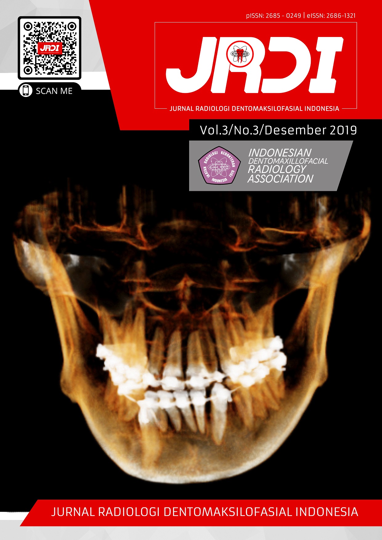
Gambaran sementoblastoma tahap awal pada cone beam computed tomography (CBCT) 3D
Abstract
Objectives: The purpose of this study was to report a case of mandibular cementoblastoma with radiologic approach and describe its characteristics.Case Report: A 32-year-old female came to the Hospital and complained of swelling of the left side of the lower jaw. Clinical examination showed a strong swelling in the buccal region of teeth 44-45, with the same soft tissue color as the surrounding tissue. In the picture cone beam computed tomography appears as a rounded lesion, the internal structure of the radiointermediet with clear and firm boundaries, surrounded by a halo radiolucent. Cementoblastoma radiodiagnosis is established. The patient was referred for surgery.
Conclusion: Cementoblastoma was a benign tumor with radiographic characteristics in the form of clearly demarcated radiopaque lesion with radiolucent halo. Some early-stage lesions can show lower density.
References
Kumar S, Angra R, Prabhakar V. Infected cementoblastoma. Natl J Maxillofac Surg. 2011;2(2):200.
Milani CM, Thomé CA, Sayuri R, et al. Mandibular cementoblastoma: Case report. Open J Stomatol. 2012;2(March):50-3.
Dogra KS, Sharma A, Sharma N, Sharma A. Cementoblastoma a Rare Odontogenic Tumor -A Case Report and Differential Diagnosis Cementoblastoma a Rare Odontogenic Tumor - A Case Report and Differential Diagnosis. 2016;(September):5-8.
4. Subramani V, Narasimhan M, Ramalingam S, Anandan S, Ranganathan S. Revisiting Cementoblastoma with a Rare Case Presentation. Case Rep Pathol. 2017;2017:1-3.
Sharma N. Benign cementoblastoma: A rare case report with review of literature. Contemp Clin Dent. 2014;5(1):92-4.
Nuvvula S, Manepalli S, Mohapatra A, Mallineni SK. Cementoblastoma Relating to Right Mandibular Second Primary Molar. Case Rep Dent. 2016;2016(Figure 2):1-5.
Huber AR, Folk GS. Cementoblastoma. Head Neck Pathol. 2009;3(2):133-5.
Mortazavi H, Baharvand M, Rahmani S, Jafari S, Parvaei P. Radiolucent rim as a possible diagnostic aid for differentiating jaw lesions. Imaging Sci Dent. 2015;45(4):253-261.
Sankari LS, Ramakrishnan K. Benign cementoblastoma. J Oral Maxillofac Pathol. 2011;15(3):358-360.
White SC, Pharoah MJ. Oral Radiology Principle and Interpretation. 7th ed. Missouri: Elsevier; 2014.
Published
2020-01-15
How to Cite
GRACEA, Rellyca Sola; FIRMAN, Ria Noerianingsih.
Gambaran sementoblastoma tahap awal pada cone beam computed tomography (CBCT) 3D.
Jurnal Radiologi Dentomaksilofasial Indonesia (JRDI), [S.l.], v. 3, n. 3, p. 49-51, jan. 2020.
ISSN 2686-1321.
Available at: <http://jurnal.pdgi.or.id/index.php/jrdi/article/view/446>. Date accessed: 10 feb. 2026.
doi: https://doi.org/10.32793/jrdi.v3i3.446.
Section
Case Report


