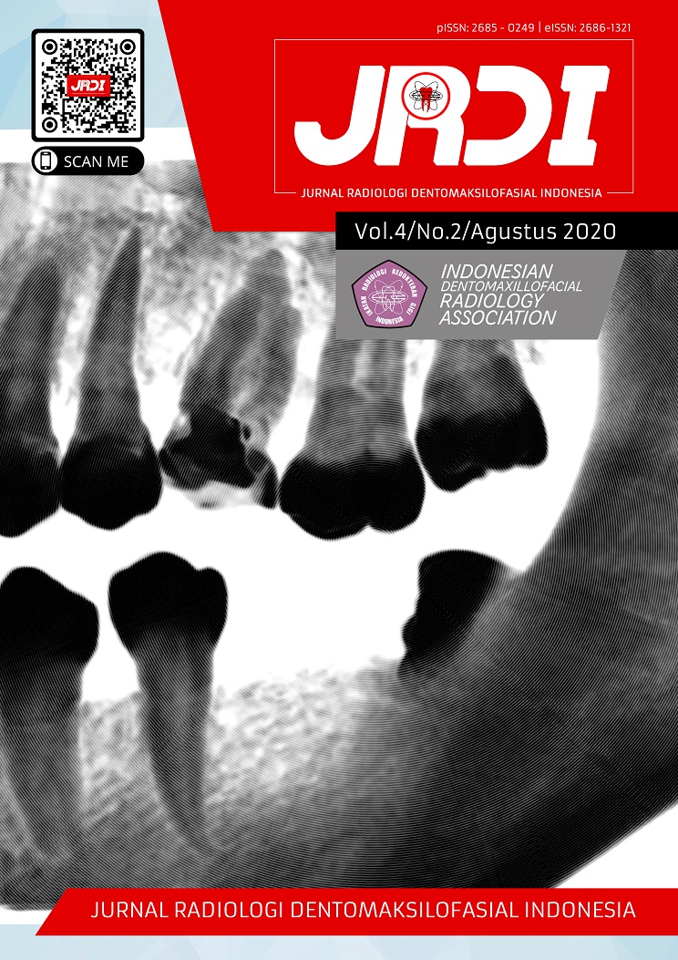
Klasifikasi impaksi caninus rahang atas pada pemeriksaan radiograf panoramik dan CBCT sebagai penunjang odontomy
Abstract
Objectives: To assess the difficulty level of dental impaction treatment. This article discusses the problem of odontomy treatment based on the classification of maxillary canine impaction through panoramic radiograph examination and CBCT.Literature Review: Impacted tooth is pathological where the tooth fails to reach its normal functional position. Impaired maxillary canine second order placement after impact of third molars. The location of impacted jaw canine teeth most often occurs in the palatal region with a horizontal position according to the maxillary sinus and nasal cavity so that complications are required during odontomy.
Conclusion: Based on this literature study, classification on impacted maxillary canine teeth has been developed based on panoramic (2D) and CBCT (3D) radiography, so thus resulting in a complete classification of impacted maxillary canine teeth and can be used as a predictor of the difficulty level of maxillary canine tooth impaction treatment.
References
Alassiry A. Radiographic assessment of the prevalence, pattern and position of maxillary canine impaction in Najran (Saudi Arabia) population using orthopantomograms – A cross-sectional,retrospective study. Saudi Dental Journal 2020; 32 : 155-9.
Al-Zoubi, H, Alharbi AA, Ferguson, DJ, Zafar MS. Frequency of impacted teeth and categorization of impacted canines: a retrospective radiographic study using orthopantomograms. Eur. J. Dent 2017 ; 11 : 117–121.
Alqerban A, Jacobs R, Fieuws S,Willem G. Comparison of two cone beam computed tomographic systems versus panoramic imaging for localization of impacted maxillary canines and detection of root resorption. The European Journal of Orthodontics 2011 ; 33(1) : 93-102.
Becker A, Chaushu S, Casap-Caspi N. Cone-beam computed tomography and the orthosurgical management of impacted teeth. Journal of the American Dental Association 2010 ; 141
Dalessandri D, Migliorati M, Rubiano R, Visconti L, Contardo L, Di Lenarda R, et al. Reliability of a novel CBCT-based 3D classification system for maxillary canine impactions in orthodontics: the KPG index. Scientific World Journal 2013;2013:921234.
Dalessandri D, Migliorati M, Visconti L, Contardo L, Kau CH, Martin C. KPG index versus OPG measurements: a comparison between 3D and 2D methods in predicting treatment duration and difficulty level for patients with impacted maxillary canines. BioMed Res Int 2014;2014:537620.
Goyal B, Munjal S, Singh S, Natt SA, Singh H. Impacted canine : An arduos task. Journal of Applied Dental and Medical Science 2018; 4:136-41.
Grisar K, Piccart F, Rimawi A, Basso I, Politis C, Jacobs R. Three‐dimensional position of impacted maxillary canines: Prevalence, associated pathology and introduction to a new classification system. Clin Exp Dent Res 2019 ; 5 : 19-25.
Ghoneima a, kanomi R, Deguchi T. Position and Distribution of Maxillary Displaced Canine in a Japanese Population: a Retrospective Study of 287 CBCT Scans. Anat Physiol 2014 ; 4 :1-5.
Haralur SB, Al Shahrani, S Alqahtani, F Nusair, Y Alshammari, O Alshenqety. Incidence of impacted maxillary canine teeth in Saudi Arabian subpopulation at central Saudi Arabian region. Ann Trop Med 2017 ; 10 : 558–562.
Hussein MA, Watted N, Hussien E, Maxillary Impacted canines;clinical review. Int J dent and Med Science Reserch 2017 ; 1(6) :10-26
Hou, R. Investigation of impacted permanent teeth except the third molar in Chinese patients through an X–ray study. J. Oral Maxillofac Surg 2010 ; 68 : 762–767.
J Brown, R Jacobs, E Levring Jaghagen . Basic training requirements for the use of dental CBCT by dentists: a position paper prepared by the European Academy of DentoMaxilloFacial Radiology. Dentomaxillofacial Radiology 2014 ; 43 (1).
Jung Y H, Liang H, Benson BW, Flint DJ, Cho BH. The assessment of impacted maxillary canine position with panoramic radiography and cone beam CT. Dentomaxillofac Radiol 2012 ; 41 : 356– 360.
Kau CH, Lee JJ, Souccar NM, The validation of a novel index assessing canine impactions. Eur J Dent 2013;7(4):399-404
Kau CH, Migliorati M, Visconti L, Contardo L, Martin C, KPG index versus OPG measurement : a comparison between 3D and 2D methods in predicting treatment duration and difficulty level for patients with impacted maxillary canines. BMRI 2014 ; 1-7.
Kau CH, Pan P, Gallerano RL, English JD, A novel 3D classification system for canine impactions--the KPG index. Int J Med Robot Comput Assist Surg MRCAS 2009;5(3):291-6.
Konda P, Ahmed MU, Ali SM, Konda A. Impacted Maxillary Canine –At a Glance.IJCD 2011; 2(6): 65-70.
Kim Y, Hyun H-K, Jang K-T. Interrelationship between the position of impacted maxillary canines and the morphology of the maxilla. Am J Orthod Dentofac Orthop Off Publ Am Assoc Orthod Its Const Soc Am Board Orthod 2012;141(5):556-62
Lai CS, Bornstein MM, MMock L, Heurbeger BM. Impacted Maxillary canines and root resorptions of neighbouring teeth : a radiographicanalysis using cone-beam computed. European Journal of Orthodontics 2013 ; 35 : 529-38.
Lindauer SJ, Rubbenstein LK, Hang WM, Andersen C, Isachoon RJ. Canine impaction identified early with panoramic radiographs. JADA 2012 ; 123 ; 92-7.
Yamamato G, Ohta Y, Tsuda Y, Tanka A, Nishikawa M, Inoda H. A new classification of impacted canines and second premolars using ortopanatomography. Asian J Oral Maxillofac Surg 2003; 15:31-7.
Published
2020-08-31
How to Cite
RACHMAWATI, Ika; FIRMAN, Ria Noerianingsih.
Klasifikasi impaksi caninus rahang atas pada pemeriksaan radiograf panoramik dan CBCT sebagai penunjang odontomy.
Jurnal Radiologi Dentomaksilofasial Indonesia (JRDI), [S.l.], v. 4, n. 2, p. 35-42, aug. 2020.
ISSN 2686-1321.
Available at: <http://jurnal.pdgi.or.id/index.php/jrdi/article/view/532>. Date accessed: 25 feb. 2026.
doi: https://doi.org/10.32793/jrdi.v4i2.532.
Section
Review Article

This work is licensed under a Creative Commons Attribution-NonCommercial-NoDerivatives 4.0 International License.


