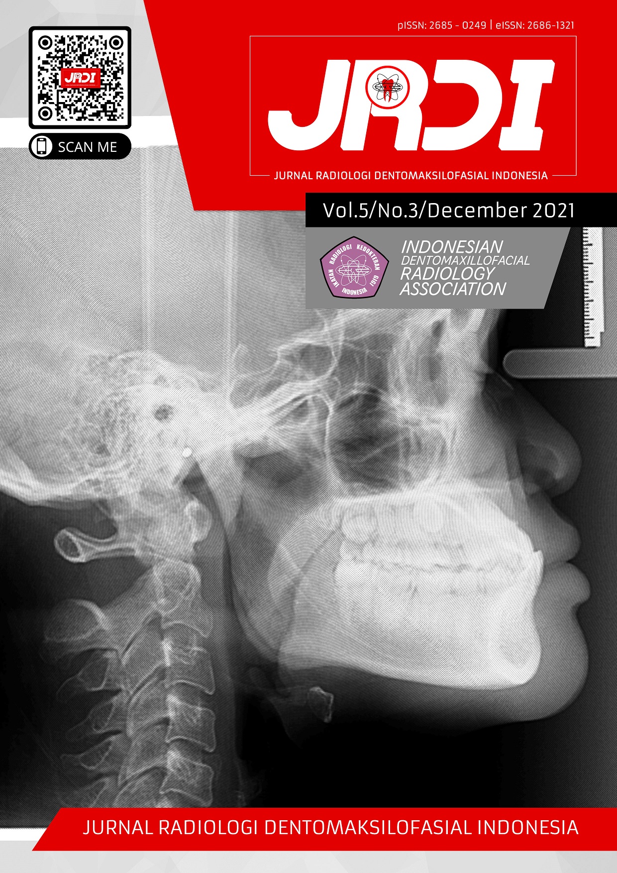
Progressive systemic sclerosis with unilateral osteolysis of the mandible: a unique case report and review
Abstract
Objectives: This case report is aimed to discuss case findings of Progressive Systemic Sclerosis (PSS), an overview of the characteristics in the form of osteolysis on one side of the mandible, and a theoretical review.Case Report: A 30-year old male patient came to an oral surgeon after tooth extraction. Clinical extraoral examination revealed hyperpigmentation on the right side of the face. A radiological study showed widening periodontal space on posterior teeth, and the angles of the mandibular arch, the jaw branch and the mandibular condyle neck were dissolved in the form of bone resorption.
Conclusion: Characteristics of Progressive Systemic Sclerosis (PSS) in radiographs appear in the form of expansion of the periodontal space and osteolysis of the mandibular angle, branch, and even condyle. This disease is caused by an autoimmune disease that affects the entire body, but it can manifest on one side of the body.
References
Panchbhai A, Pawar S, Barad A. Kazi Z. Review of orofacial considerations of systemic sclerosis or scleroderma with report of analysis of 3 cases. Indian J Dent. 2016;7(3):134-9.
Sharma ML, Mehrotra G, Sharma K, Suman S. Tail of the whale appearance: A pathognomonic feature of scleroderma. Journal of Indian Academy of Oral Medicine & Radiology. 2020; 32(1):55-9.
Srivastava R, Jyoti B, Bihari M, Pradhan S. Progressive systemic sclerosis with intraoral manifestations: A case report and review. Indian J Dent. 2016;7(2):99-104.
Marcucci M, Abdala A. Clinical and radiographic study of orofacial alterations in patients with systemic sclerosis. Brazil Oral Reserch. 2009;23(1):82-8.
Hasan S, Khan R,Haroon MA,Jahan Khwaja K, Tarannum F,Hussain AR. Systemic Sclerosis-A Case Report and Review of Literature. Int. Journal of Contemporary Dentistry. 2011;2(6):46-53.
Anbiaee, N, Tafakhori Z. Early diagnosis of progressive systemic sclerosis (scleroderma) from a panoramic view: report of three cases. Dentomaxillofacial Radiology. 2011;40:457-62.
White SC, Pharoah MJ, editors. Oral radiology principles and interpretation, 7th edition. Saint Louis: Mosby, 2014: p.465-7.
Chung L, Lin J, Furst E, Florentino D. Systemic and localized scleroderma. Clin Dermatol. 2006;24:374-92.
Dokumen pasien. RSGM Universitas Hang Tuah Surabaya, 2020.
Sharma ML, Mehrotra G, Sharma K, Suman S. Tail of the whale appearance: A pathognomonic feature of scleroderma. Journal of Indian Academy of Oral Medicine & Radiology. 2020;32:55-9.
Bali V, Dabra S, Behl AB, Bali R. A rare case of hidebound disease with dental implications. Dent Res J (Isfahan) 2013;10(4):556‑61.
Veale B, Jablonski RY, Frech TM, Pauling JD. Orofacial manifestations of systemic sclerosis. British Dental Journal. 2016;221(6):305-10.
Leader D, Papas A, Finkelman M. A survey of dentists’ knowledge and attitudes with respect to the treatment of scleroderma patients. J Clin Rheumatol. 2014;20:189–94.
Mehra A. Periodontal space widening in patients with systemic sclerosis: a probable explanation. Dentomaxillofac Radiol. 2008;37:183.
de Figueiredo MAZ, de Figueiredo JAP, Porter S. Root resorption associated with mandibular bone erosion in a patient with scleroderma. Journal Endodontic. 2008;34:102‑3.
Hasan S, Khan R, Haroon MA, Khwaja KJ, Tarannum F, Hussain AR. Systemic sclerosis: a case report and review of literature. Int J Contemp Dent. 2011;2:46‑53.
Nortje CJ. Case report: Maxillofacial radiology case 117. The South African Dental Journal. 2014;69(1):25.
Rahpeyma A, Zarch SHH, Khajehahmadi S. Severe osteolysis of the mandibular angle and total condylolysis in progressive systemic sclerosis. Case Reports in Dentistry. 2013:948042.
Haers PE, Sailer HF. Mandibular resorption due to systemic sclerosis. Case report of surgical correction of a secondary open bite deformity. Int J Oral Maxillofac Surg. 1995;24:261-7.
Hopper FE, Giles AD. Orofacial changes in systemic sclerosis‑report of a case of resorption of mandibular angles and zygomatic arches. Br J Oral Surg. 1982;20:129-34.
Published
2021-12-31
How to Cite
EPSILAWATI, Lusi; MEDIKA, Chrisna Ardhya; HERMANTO, Eddy.
Progressive systemic sclerosis with unilateral osteolysis of the mandible: a unique case report and review.
Jurnal Radiologi Dentomaksilofasial Indonesia (JRDI), [S.l.], v. 5, n. 3, p. 126-129, dec. 2021.
ISSN 2686-1321.
Available at: <http://jurnal.pdgi.or.id/index.php/jrdi/article/view/734>. Date accessed: 25 feb. 2026.
doi: https://doi.org/10.32793/jrdi.v5i3.734.
Section
Case Report

This work is licensed under a Creative Commons Attribution-NonCommercial-NoDerivatives 4.0 International License.


