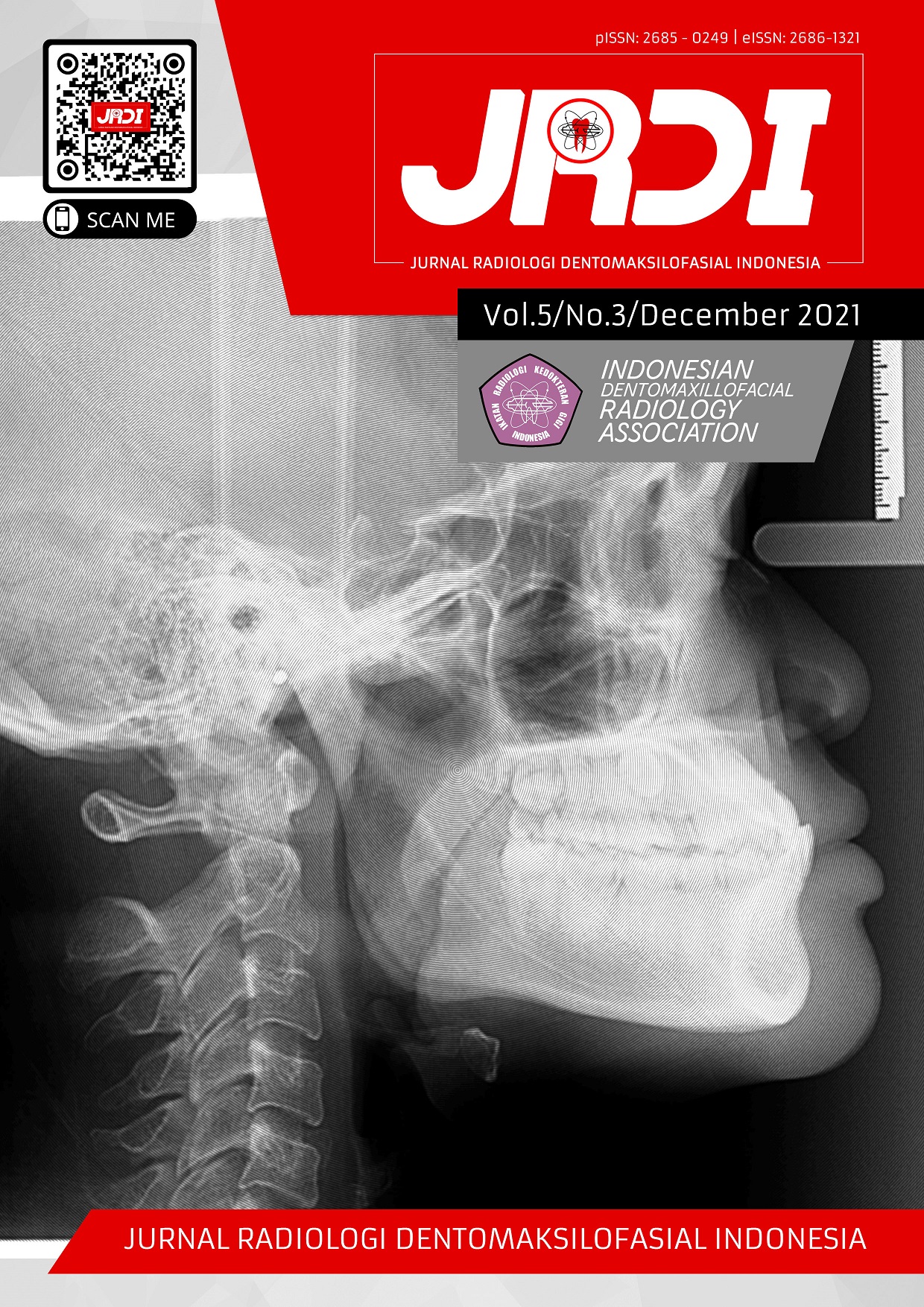
Analysis of osteoblast, osteoclast levels and radiographic patterns in the healing process of bone fractures (preliminary research)
Abstract
Objectives: The healing process of a bone fracture goes through many phases. The hard callus phase was critical where the original structure was conducted. The hard callus growth depends on osteoblasts and osteoclasts active, and this condition can be analyzed on the radiograph. This study aimed to examine the analysis of bone fracture healing between osteoblasts and osteoclast numbers and radiographic patterns.Materials and Methods: The study used 12 male Wistar rats with an incomplete fracture in the right femur made by a dental tapered bur with 0.3 mm in length and 0.2 mm in depth. Digital radiographic examinations were carried out on days 0, 5, 10, 17, and 25 after fracturing in a lateral position. Furthermore, a radiographic analysis was performed using Image-J to obtain changes in the value of length and depth in the healing area. The research was conducted to find the radiopaque and radiolucent patterns and the number of osteoblasts and osteoclasts.
Results: This study resulted in a change in the radiograph pattern. Callus formation resulted in fracture areas with a smaller distance from day 0 to day 25. The bone healing process begins with granulation tissue formation, followed by the gradual replacement of the connective tissue and bone. This process is comparable to the increase in osteoblasts up to day 25, which blocks bone resorption. Osteoclasts regulate bone resorption, and their number increases after 10 and 17 days to replace bone formation. Osteoclasts decline after 25 days because osteoblasts inhibit them, which control bone formation.
Conclusion: The conclusions were obtained there are changes in the radiograph pattern. The radiopaque increased while the radiolucent decreased; the osteoclast pattern tended to be stable and lowered while the osteoblasts increased during the fracture healing process. The correlation of all the factors is very closely related.
References
Oryan A, Monazzah S, Bigham-Sadegh A. Bone injury and fracture healing biology. Biomed Environ Sci. 2015;28(1):57–71.
Fikri M, Azhari A, Epsilawati L. Gambaran kualitas tulang pada wanita berdasarkan kelompok usia melalui radiografi panoramik. J Radiol Dentomaksilofasial Indones. 2020;4(2):5.
Mock C, Cherian MN. The global burden of musculoskeletal injuries: Challenges and solutions. Clin Orthop Relat Res. 2008;466(10):2306–16.
Riskesdas. Riset Kesehatan Dasar [Internet]. Jakarta; 2018. Available from: http://www.depkes.go.id/resources/download/info-terkini/materi_rakorpop_2018/Hasil Riskesdas 2018.pdf
Kovtun A, Bergdolt S, Wiegner R, Radermacher P, Huber-Lang M, Ignatius A. The crucial role of neutrophil granulocytes in bone fracture healing. Eur Cells Mater. 2016;32(Cxcl):152–62.
Einhorn TA, Gerstenfeld LC. Fracture healing: mechanisms and interventions Thomas. Nat Rev Rheumatol. 2015;11(1):45–54.
Sabri M. Administration’s Effects of Ethanol Extract of Cissus quadrangularis Salisb on Growth of Lumbal Bone in Ovariectomized Rats. Natural. 2013;13(2):48–54.
Jayakumar P, Di Silvio L. Osteoblasts in bone tissue engineering. Proc Inst Mech Eng Part H J Eng Med. 2010;224(12):1415–40.
Blokhuis TJ, De Bruine JHD, Bramer JAM, Den Boer FC, Bakker FC, Patka P, et al. The reliability of plain radiography in experimental fracture healing. Skeletal Radiol. 2001;30(3):151–6.
Gunawan G, Sitam S, Epsilawati L. Densitas tulang mandibula pengguna obat anti hipertensi calcium channel blocker (CCB) melalui radiograf panoramik. J Radiol Dentomaksilofasial Indones. 2020;4(2):1.
Epsilawati L, Satari M, Azhari. Analysis of Myrmecodia Pendens in Bone Healing Process to Improve the Quality of Life: Literature Review. IOP Conf Ser Earth Environ Sci. 2019;248(1).
Chen WT, Han DC, Zhang PX, Han N, Kou YH, Yin XF, et al. A special healing pattern in stable metaphyseal fractures. Acta Orthop. 2015;86(2):238–42.
Ghiasi MS, Chen J, Vaziri A, Rodriguez EK, Nazarian A. Bone fracture healing in mechanobiological modeling: A review of principles and methods. Bone Reports [Internet]. 2017;6:87–100.
Haffner-Luntzer M, Fischer V, Prystaz K, Liedert A, Ignatius A. The inflammatory phase of fracture healing is influenced by oestrogen status in mice. Eur J Med Res. 2017;22(1):1–11.
Sathyendra V, Darowish M. Basic science of bone healing. Hand Clin [Internet]. 2013;29(4):473–81.
Utomo DN. Defek Kartilago Sendi Lutus. Surabaya: AIRLANGGA Unversity Press; 2018.
Lerner UH. Osteoblasts, Osteoclasts, and Osteocytes: Unveiling Their Intimate-Associated Responses to Applied Orthodontic Forces. Semin Orthod [Internet]. 2012;18(4):237–48.
Buza JA, Einhorn T. Bone healing in 2016. Clin Cases Miner Bone Metab. 2016;13(2):101–5.
Eastaugh-Waring SJ, Joslin CC, Hardy JRW, Cunningham JL. Quantification of fracture healing from radiographs using the maximum callus index. Clin Orthop Relat Res. 2009;467(8):1986–91.
Salih S, Blakey C, Chan D, McGregor-Riley JC, Royston SL, Gowlett S, et al. The callus fracture sign: a radiological predictor of progression to hypertrophic non-union in diaphyseal tibial fractures. Strateg Trauma Limb Reconstr. 2015;10(3):149–53.
Published
2021-12-31
How to Cite
SARIFAH, Norlaila et al.
Analysis of osteoblast, osteoclast levels and radiographic patterns in the healing process of bone fractures (preliminary research).
Jurnal Radiologi Dentomaksilofasial Indonesia (JRDI), [S.l.], v. 5, n. 3, p. 106-111, dec. 2021.
ISSN 2686-1321.
Available at: <http://jurnal.pdgi.or.id/index.php/jrdi/article/view/740>. Date accessed: 25 feb. 2026.
doi: https://doi.org/10.32793/jrdi.v5i3.740.
Section
Original Research Article

This work is licensed under a Creative Commons Attribution-NonCommercial-NoDerivatives 4.0 International License.


