Radiographic imagery of aggressive plexiform-type ameloblastoma in the mandible: a case report
A rare case report
Abstract
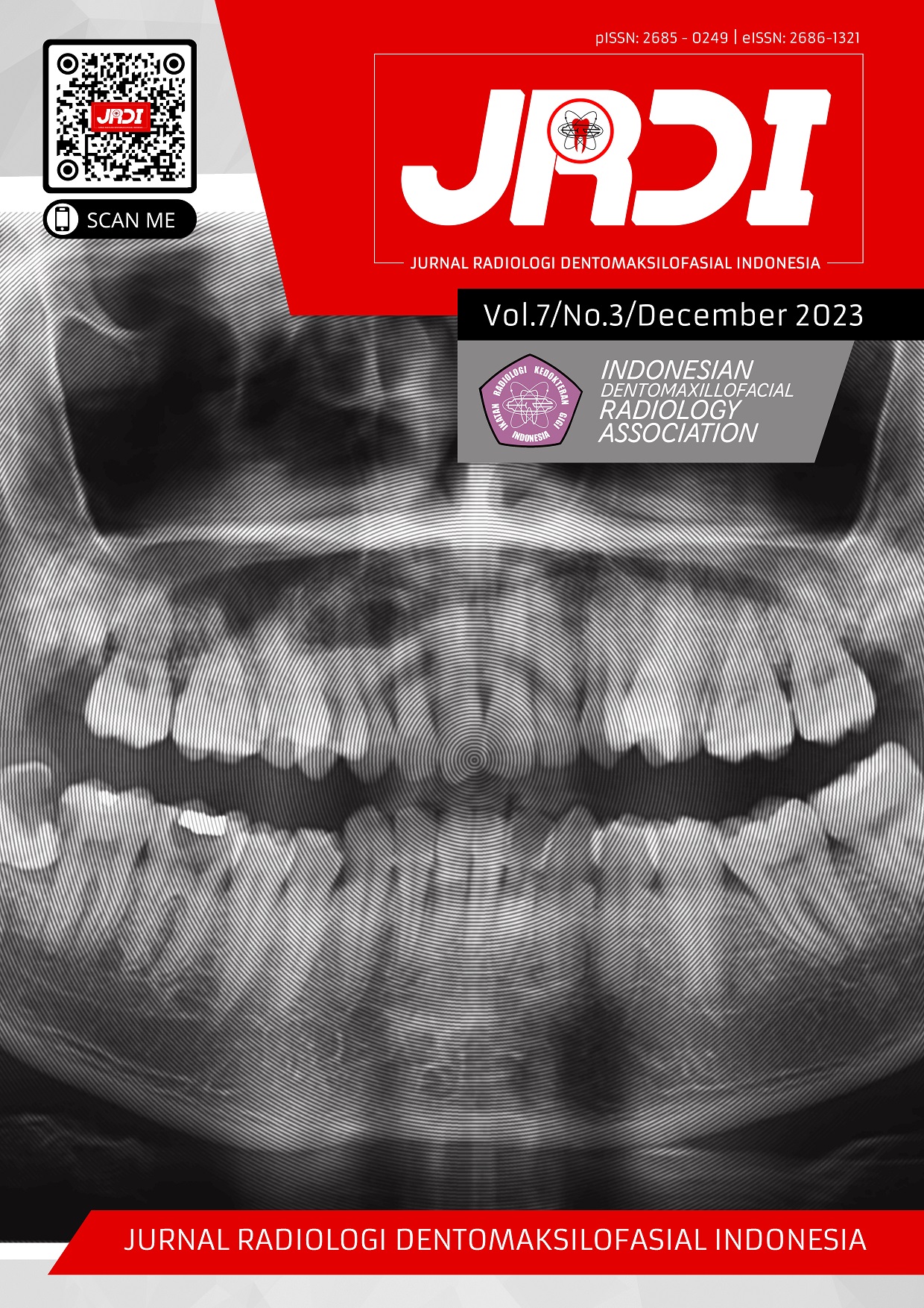
Objectives: This case report aims to describe the radiographic characteristics of plexiform ameloblastoma and its impact on surrounding tissues in a middle-aged female patient using panoramic radiography and CBCT, along with case management.Case Report: A 43-year-old female patient presented to the Dental Radiology Unit of RSGMP UNHAS with a panoramic referral letter, diagnosed clinically with anterior mandibular ameloblastoma. Extraoral examination revealed an asymmetrical facial appearance with anterior mandibular enlargement. Intraoral examination showed mucus membrane enlargement in the anterior mandible region (teeth 37-45), soft consistency, absence of crepitus, no palpation tenderness, and mobility in several anterior mandibular teeth. The first panoramic radiograph (March 16, 2022) exhibited a unilocular radiolucent lesion, well-defined, with scalloped margins in the anteroposterior mandibular region. The second panoramic examination (May 23, 2022) indicated a more aggressive lesion expansion, with evidence of root resorption and destruction of the inferior mandibular cortex approaching the mandibular angle. CBCT findings demonstrated a hypodense/radiolucent lesion extending anteroposteriorly, superiorly, and inferiorly, leading to displacement, root resorption, and destruction of the inferior mandibular cortex in the inferior direction.
Conclusion: Based on the characteristics and structure of the lesion observed through various radiographic examinations, a unilocular ameloblastoma was suspected. Histopathological examination confirmed the plexiform-type ameloblastoma.
References
White SC, Pharoah MJ. Oral radiology principles and interpretation. 7th ed. St. Louis, Missouri: Elsevier Mosby; 2014.
Shafer WG, Hine MK, Levy BM. Shafer’s Textbook of Oral Pathology, Eight edition. Elsevier; 2016. p. 130-2.
Bonanthaya K, Elavenil P, Manuel S, Kumar VV, Rai A. Oral and Maxillofacial Surgery for the Clinican. Singapore: Springer Nature; 2021.
Kashyap B, Reddy PS, Desai RS. Plexiform ameloblastoma mimicking a periapical lesion: A diagnostic dilemma. J Conserv Dent. 2012 Jan;15(1):84-6.
Underhill TE, Katz JO, Pope TL Jr, Dunlap CL. Radiologic findings of diseases involving the maxilla and mandible. AJR Am J Roentgenol. 1992 Aug;159(2):345-50.
Regezi JA, Sciubba JJ, Jordan RCK. 2017. Oral Pathology: Clinical Pathologic Correlations, Seventh Edition. St. Louis, Missouri: Elsiever; 2017. p. 279-80.
Odell EW. Cawson’s Essential of Oral Pathology and Oral Medicine. Elsevier; 2017. p.183-4.
Cohen MA, Hertzanu Y, Mendelsohn DB. Computed tomography in the diagnosis and treatment of mandibular ameloblastoma: report of cases. J Oral Maxillofac Surg. 1985 Oct;43(10):796-800.
Singer SR, Mupparapu M, Philipone E. Cone beam computed tomography findings in a case of plexiform ameloblastoma. Quintessence Int. 2009;40(8):627-30.
Kasat VO, Karjodkar FR, Ladda R. Plexiform unicystic ameloblastoma-a case report and review of literature. CHRISMED J Health Res. 2014;1(2):103-6.
Celur S, Babu KS. Plexiform ameloblastoma. Int J Clin Pediatr Dent. 2012 Jan;5(1):78-83.
Wu YH, Chang JY, Wang YP, Chiang CP. Langerhans cells in plexiform ameloblastoma. J Dent Sci. 2017;12(2):195-7.
Pramanik F, Epsilawati L, Lita YA, Herawati E. Analisis gambaran radiologis suspek ameloblastoma tipe solid pada radiograf CBCT 3D. Jurnal Radiologi Dentomaksilofasial Indonesia. 2019;3(2):15-20.
Brown NA, Betz BL. Ameloblastoma: A Review of Recent Molecular Pathogenetic Discoveries. Biomark Cancer. 2015;7(Suppl 2):19-24.
Effiom OA, Ogundana OM, Akinshipo AO, Akintoye SO. Ameloblastoma: current etiopathological concepts and management. Oral Dis. 2018;24(3):307-16.
Hertog D, Bloemena E, Aartman IH, van-der-Waal I. Histopathology of ameloblastoma of the jaws; some critical observations based on a 40 years single institution experience. Med Oral Patol Oral Cir Bucal. 2012;17(1):e76-82.
Published
2023-12-31
How to Cite
ARNAWANSAH, Rakhmat Putra Guru et al.
Radiographic imagery of aggressive plexiform-type ameloblastoma in the mandible: a case report.
Jurnal Radiologi Dentomaksilofasial Indonesia (JRDI), [S.l.], v. 7, n. 3, p. 111-116, dec. 2023.
ISSN 2686-1321.
Available at: <http://jurnal.pdgi.or.id/index.php/jrdi/article/view/954>. Date accessed: 25 feb. 2026.
doi: https://doi.org/10.32793/jrdi.v7i3.954.
Section
Case Report

This work is licensed under a Creative Commons Attribution-NonCommercial-NoDerivatives 4.0 International License.


